4038
Value of MR placental protusion sign in predicting postpartumhemorrhage in patients with placenta previa1Department of Radiology, First Hospital of Jilin University, Chang, China
Synopsis
Objective: To explore the MRI findings of placental protusion sign in predicting postpartum hemorrhage in pa—tients with placenta previa.
Methods: Totally 35 4 placenta previa patients with whole clinical data underwent MR scaning 2weeks before operation.Association of postpartum hemorrhage and placental protusion sign was analyzed.
Results: Among354 patients with placental previa,the age of the pregnant women(#一4.34,P一0.04),gestational age at delivery(z2—5.19,P一0.02)and the number of cesarean sections(Z2=44.85,P<O.01)had associated with postpartum hemorrhage.Eight cases had placental protusion sign in MRI,while 6 cases occurred postpartum hemorrhage.The incidence of postpar—tum hemorrhage was 75.00%(6/8)and 12.72%(44/346)in patients with placenta accreta and with placental abruption。respectively(3[2—20.14,P<O.01).The sensitivity,specificity,odds ratio(95%confidence interval)and positive likeli—hood ratio of predicting postpartum hemorrhage was 12.00%(6/50),99.34%(302/304),20.59(4.03,105.23)and15.68,respectively.
Conclusion MRI placental protrusion sign has important clinical reference value in predicting postpar—tum hemorrhage.
Introduction
With the increase of cesarean section rate, postpartum hemorrhage caused by placental factors is also increasing [1]. Previous studies on placenta implantation focused on the analysis of the signs of placenta implantation and the guiding significance of pathological types, while few reports on the value of MRI signs in predicting postpartum hemorrhage. The purpose of this study was to analyze the value of MRI placental prominence in predicting postpartum hemorrhage.Methods
Totally 35 4 placenta previa patients with whole clinical data underwent MR scaning 2weeks before operation.Association of postpartum hemorrhage and placental protusion sign was analyzed.SPSS 17 statistical analysis software was used. The sensitivity, specificity, odds ratio (OR) and positive likeli hood ratio (+ LR) of placental herniation in the diagnosis of postpartum hemorrhage were calculated and compared with Y2 test. P<0.05 was statistically significant.Results
Among354 patients with placental previa,the age of the pregnant women(#一4.34,P一0.04),gestational age at delivery(z2—5.19,P一0.02)and the number of cesarean sections(Z2=44.85,P<O.01)had associated with postpartum hemorrhage.Eight cases had placental protusion sign in MRI,while 6 cases occurred postpartum hemorrhage.The incidence of postpar—tum hemorrhage was 75.00%(6/8)and 12.72%(44/346)in patients with placenta accreta and with placental abruption。respectively(3[2—20.14,P<O.01).The sensitivity,specificity,odds ratio(95%confidence interval)and positive likeli—hood ratio of predicting postpartum hemorrhage was 12.00%(6/50),99.34%(302/304),20.59(4.03,105.23)and15.68,respectivelyDiscussion
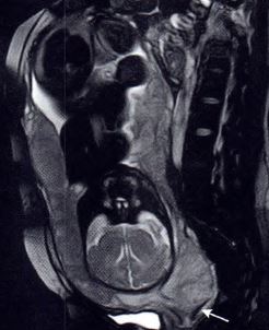
Placental factors leading to postpartum hemorrhage mainly include abnormal placental attachment and abnormal attachment.[2] Postpartum hemorrhage mainly occurs in abnormal placenta position, which is closely related to the tissue structure. The muscle tissue of lower uterine segment is thin and its contractility is poor. When the placenta attaches to this area, it is difficult to completely dissect the placenta, and postpartum hemorrhage often occurs. The abnormal attachment of placenta mainly includesPlacenta adhesion, placenta implantation and placental penetration are 3 types. This study focuses on a special condition of placental position, namely, the placental process of human cervix (Figure 1).Conclusion
MRI placental protrusion sign has important clinical reference value in predicting postpar—tum hemorrhage.Acknowledgements
None.References
[1] Sannananja B, Ellermeier A, Hippe DS, et al. Utility of diffusion-weighted MR imaging in the diagnosis of placenta accreta spectrum abnormality. Abdom Radiol (NY). 2018 Nov;43(11):3147-3156.
[2]Ghaghada KB, Starosolski ZA, Bhayana S, et al. Pre-clinical evaluation of a nanoparticle-based blood-pool contrast agent for MR imaging of the placenta. Placenta. 2017 Sep;57:60-70.
Figures