4037
Potential utility of T1 rho imaging for diagnosis of cystic tumorHiroyuki Morisaka1, Katsuhiro Sano1, Taiki Seno1, Yasuo Sakurai1, and Tomoaki Ichikawa1
1Diagnostic Radiology, Saitama Medical Center International Medical Center, Saitama, Japan
Synopsis
T1 rho imaging is sensitive to the tissue macromolecular inclusion such as proteoglycan of meniscus cartilage. In this study, we demonstrated T1 rho values were differ in ovarian cystic tumors and not in hepatic solid tumors.
Target audience
This presentation will be targeted to audience interested in clinical application of T1 rho imaging for the diagnosis of cystic and solid neoplasm.Purpose
In the imaging diagnosis of cystic tumors such as ovarian neoplasms, an appearance of cystic component is a valuable information for the precise diagnosis, however, which is usually assessed only qualitatively. In the pathological diagnosis, pathologists diagnose cystic tumors mainly based on the epithelial character not on fluid component. T1 rho imaging, which is sensitive to the tissue macromolecular inclusion, is introduced in a clinical MR imaging for assessing meniscus cartilage of the knee joint and liver and myocardial fibrosis1)2). We speculate T1 rho imaging would be useful for tumor diagnosis by providing some important clues about tumor tissue macromolecular characteristics.Methods
Surgically resected and pathologically confirmed 28 cases of ovarian cystic tumors (10 cases of serous cystic tumors, 14 cases of mucinous cystic tumors, and 4 cases of endometrioid tumors, mean tumor size is 124±50 mm) and 47 cases of solid hepatic cancers (25 cases of hepatic metastasis and 22 cases of hepatocellular carcinoma, mean tumor size is 42±30 mm) were included in this retrospective study, for all of which preoperative MR imaging including single slice T1 rho imaging and diffusion weighted imaging was performed. In T1 rho imaging, single slice was set at the maximum cross-sectional area of the tumor, and the spin lock pulse amplitude was set at 500 Hz and the TSLs were 1, 20, 40, and 60 ms. T1ρ maps were generated on a pixel-by-pixel basis using a mono-exponential decay model. By placing a region of interest (ROI) on the target lesion on an axial T1 rho map, T1 rho values [ms] of cystic component of ovarian tumors and solid component of hepatic malignancies were calculated. Apparent diffusion coefficient (ADC) map was generated using axial diffusion weighted images with b-values of 0 and 1000 s/mm2, and ADC values of the same ROI at the same imaging plane with the T1 rho map were calculated. T1 rho values and ADC values [mm2/s] were compared by Wilcoxon or Kruskal-Wallis test.Results
There was a significant difference in T1 rho values among the three types of ovarian tumors (p<0.01, Figure 1 and 2), while there was no significant difference in those between the two types of hepatic cancers. ADC value showed no significant group difference neither in the ovarian cystic tumors nor hepatic cancers.Discussion
Cystic and solid component of tumors include various types of macromolecules, which would be vary from one entity to another. From our results, T1 rho imaging would be sensitive to the difference of tumor fluid content and not sensitive to that of solid component.Conclusion
T1 rho imaging will be sensitive to the difference of fluid component and has a potential utility for the diagnosis of cystic tumors.Acknowledgements
No acknowledgement found.References
- Takayama Y, Nishie A, Asayama Y, et al. T1 rho Relaxation of the liver: A potential biomarker of liver function. J Magn Reson Imaging. Jul 2015;42(1):188-195.
- Wang L, Regatte RR. T(1)rho MRI of human musculoskeletal system. J Magn Reson Imaging. Mar 2015;41(3):586-600.
Figures
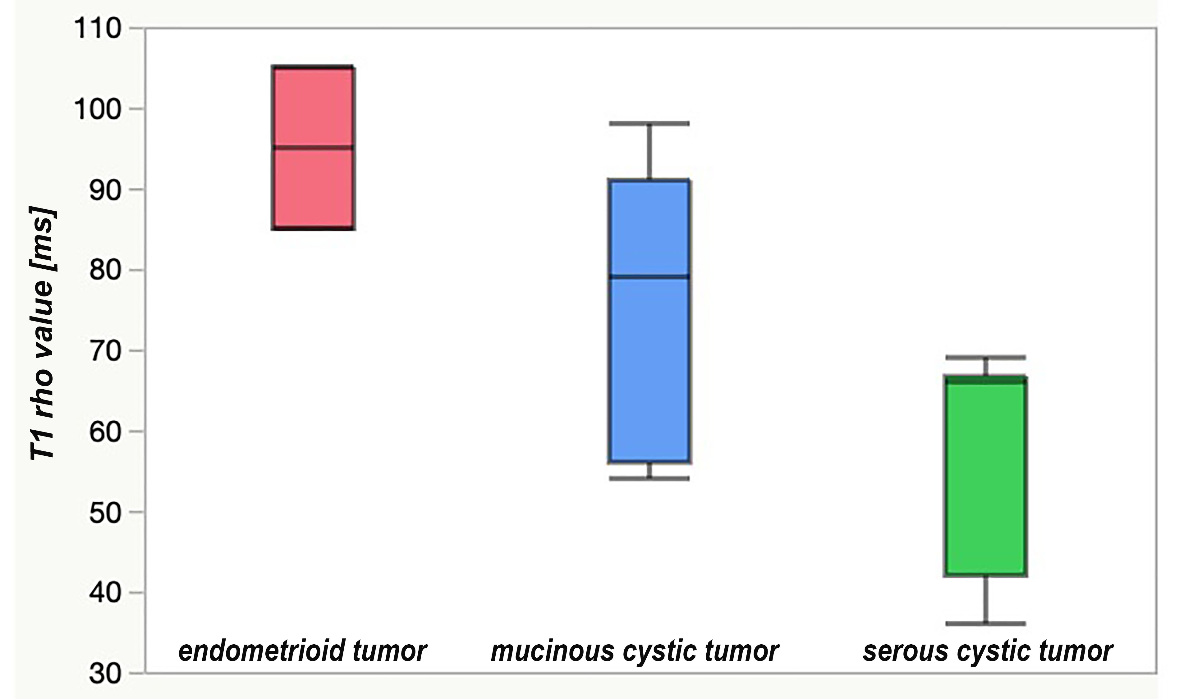
Figure 1: T1 rho values of three types of ovarian cystic tumors
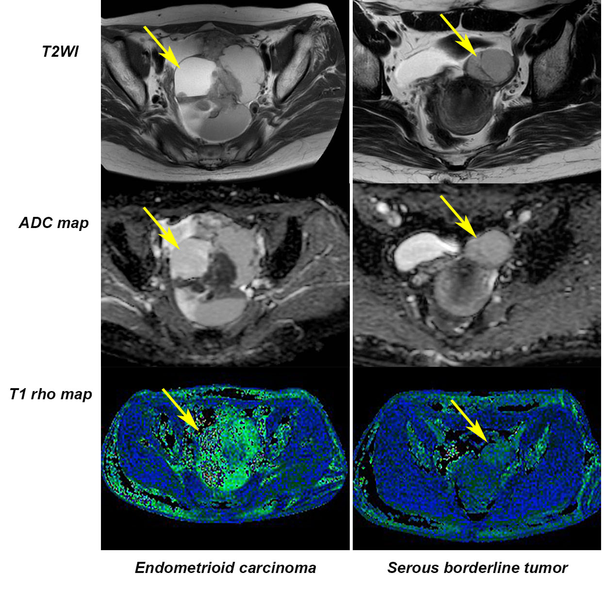
Figure 2: Examples of ovarian tumors with a high and low T1 rho values