4029
Achieving Real-Time QSM Reconstruction Using Deep Neural Network1Department of Electrical and Computer Engineering, Seoul National University, Seoul, Korea, Republic of, 2AIRS medical, Seoul, Korea, Republic of
Synopsis
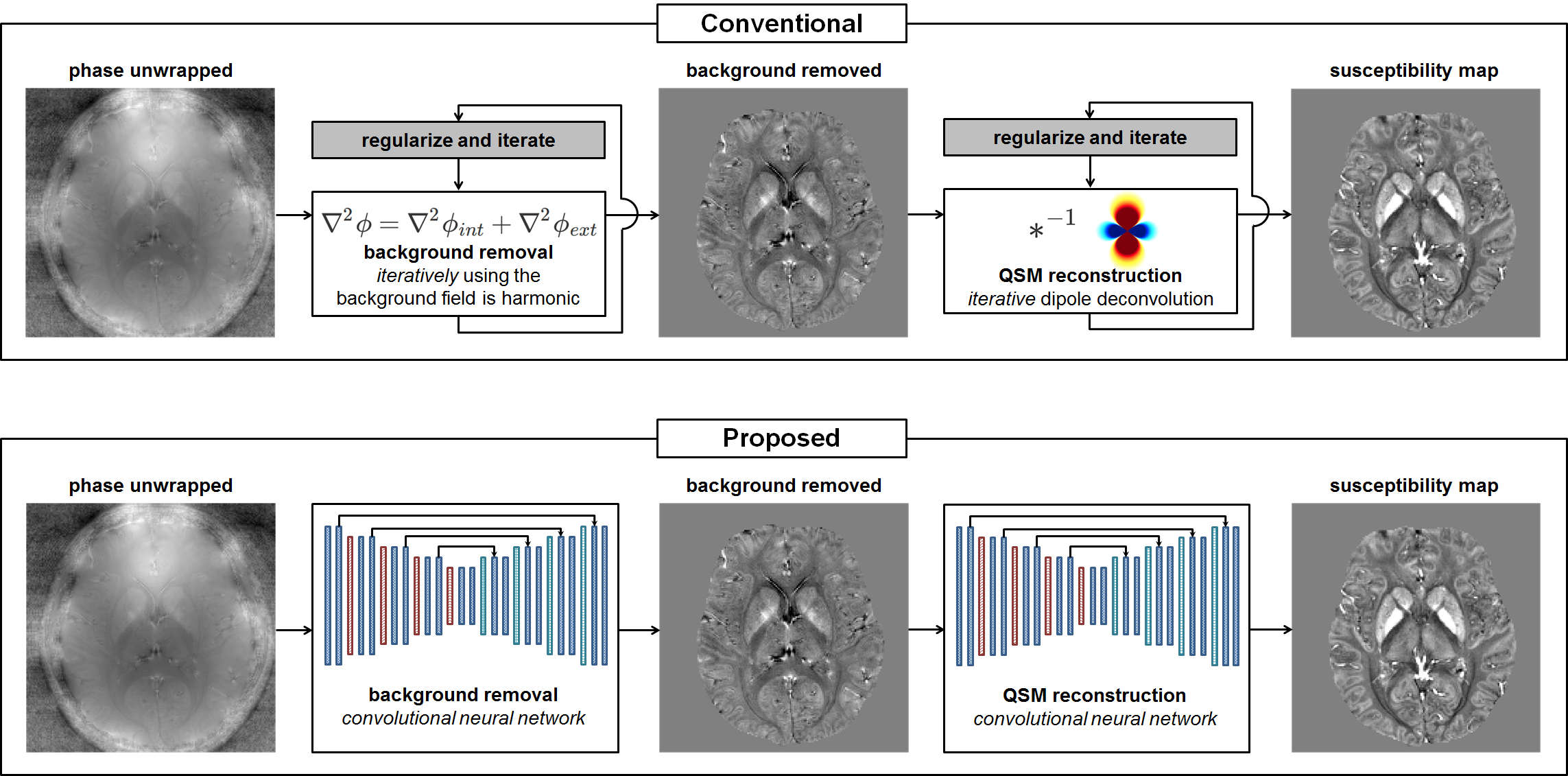
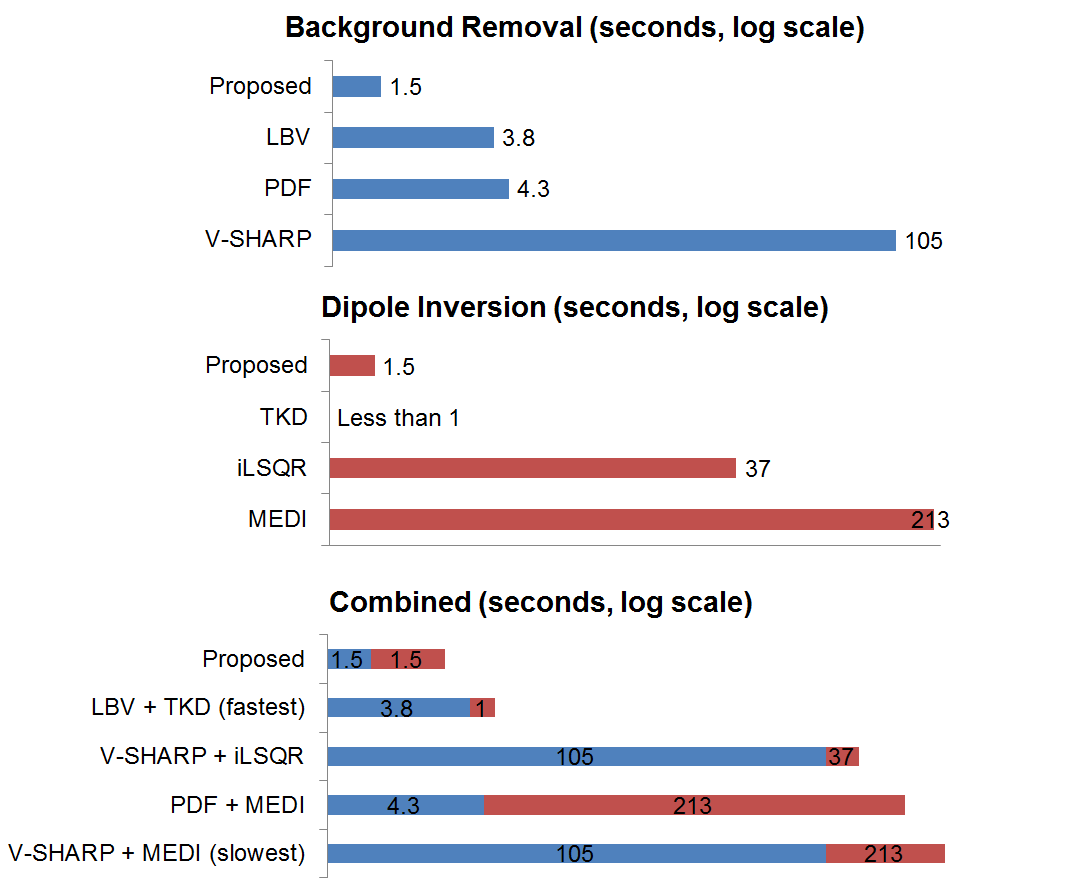
Conventional QSM reconstruction algorithms impose long computation time, which inhibits their adoption for real-time clinical use. In this work, we propose a method that replaces conventional iterative algorithms for background removal and dipole inversion with two deep neural networks. The reconstruction results demonstrate comparable performance to the previous outcomes while the new method takes only 3 seconds (up to 106 times faster!), which is unparalleled to conventional methods.
Purpose
Quantitative susceptibility mapping (QSM) has gained much attention due to its potential for the detection and quantification of certain pathological conditions such as hemorrhage, calcification and demyelination. However, conventional model-based QSM reconstruction algorithms are computationally expensive, taking several minutes of reconstruction time. Therefore, applying them for routine clinical use is challenging. In this work, we resolve this issue by replacing the background removal step and the dipole inversion step with deep neural networks.Methods
[Background Removal]
A modified U-net for 3D data processing was utilized for the background removal step. The datasets for training and test were from QSMnet1. For the training of the network, total fifteen 3D GRE phase images from three subjects (five head orientations each) were used. When training, a Laplacian-based phase unwrapped image was used as the input and a local field map obtained by V-SHARP2 was used as the output. The image was split into 64 × 64 × 64 patches with a 75% overlap, resulting in the total number of 17,160 patches. L1 loss was used as the cost function and was optimized using Adam with the learning rate of 10-5. The network was implemented using TensorFlow and was trained for 18 hours with one NVIDIA 1080Ti GPU.
[Dipole Inversion]
The dipole inversion was performed using QSMnet, which paired local field maps with gold-standard COSMOS3 maps. Detailed methods can be found in the paper1.
[Evaluation]
For the evaluation, the 3D phase images from six subjects with five head orientations each (total thirty images) were processed for Laplacian-based phase unwrapping. Then the results were fed into the background removal network. The output data from the background removal network were fed directly into the dipole inversion network, generating the final QSM map.
In order to compare the performance of the combined neural network with multiple different combinations of conventional background removal (LBV4, PDF5 and V-SHARP) and dipole inversion (TKD6, iLSQR7 and MEDI8) algorithms, the same test sets were applied. The quality of the resulting QSM maps in terms of NRMSE, SSIM, HFEN and PSNR was evaluated. The data processing times were measured for all the methods using one NVIDIA GeForce 1080Ti GPU for the proposed network and Intel i5-6600 CPU (4 cores) @ 3.30GHz for the rest. In addition, reconstructed images from each algorithm were visually scrutinized for artifacts and structural losses.
Results
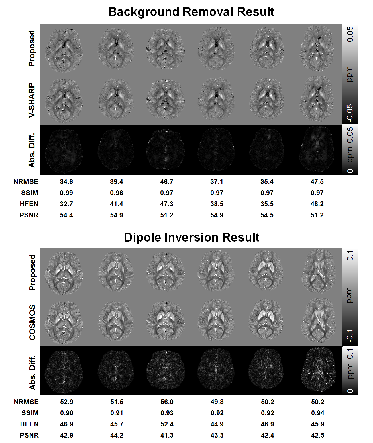
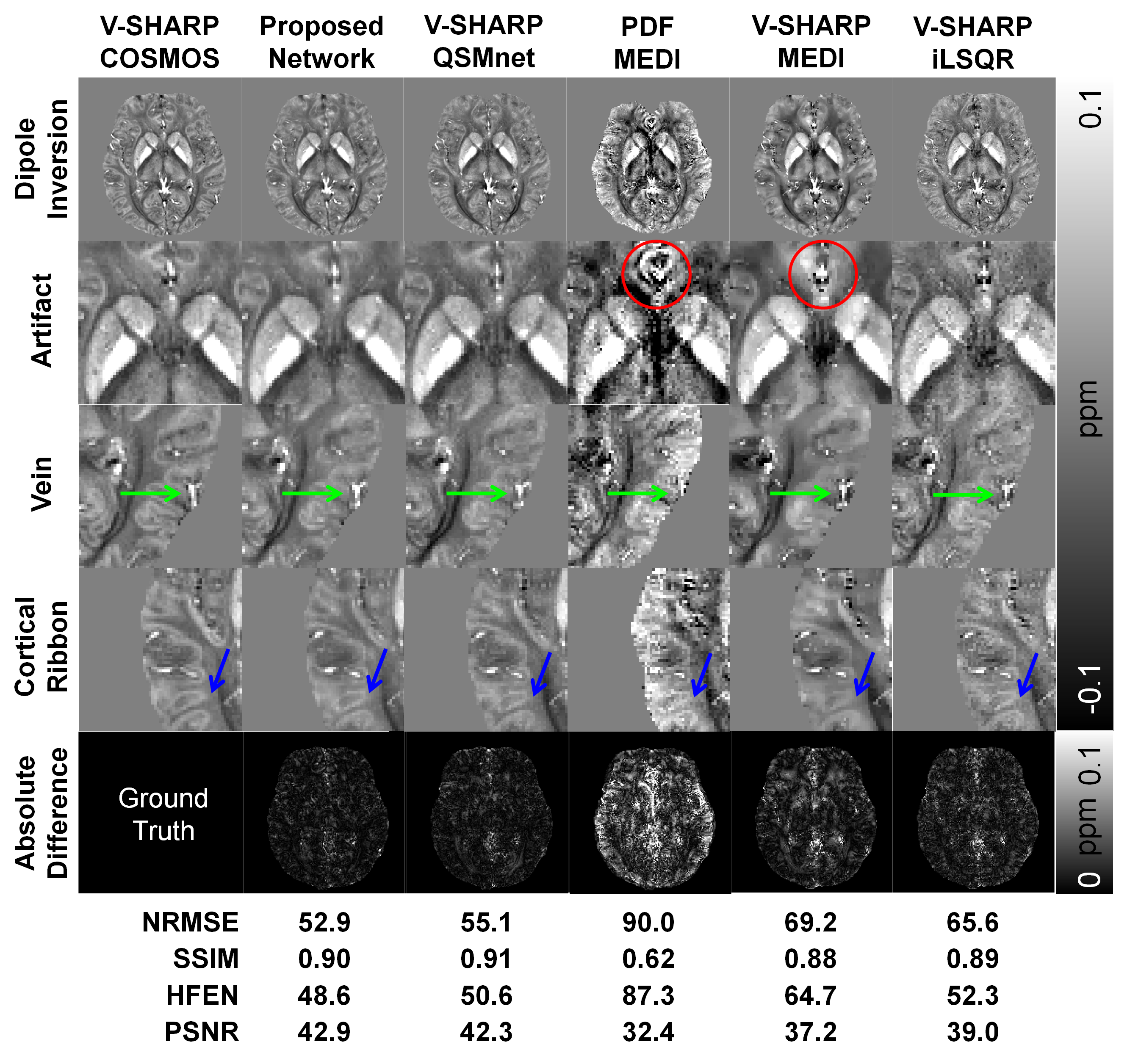
When evaluated, both networks perform nicely with satisfying results as shown in Figure 2. The NRMSE errors in the background removal network (mean NRMSE: 40.1±5.7) were lower than those of the dipole inversion network (mean NRMSE: 51.8±2.4). The other quantitative metrics showed the same trend. When the processing times were compared, the combined network (3 sec) is substantially faster (up to ×106) than the combinations of the conventional algorithms (Figure 3). The newly proposed background removal network was faster than all the other methods by a factor of 2.5 to 70 (upper row in Figure 3). Figure 4 compares the image quality of the conventional QSM methods and the combined network QSM results. The proposed network with QSMnet had the least error as shown by the error map and in terms of NRMSE, SSIM, HFEN and PSNR. Structural details such as veins and cortical ribbons were mostly preserved when reconstructed using the proposed method. On the other hand, other methods suffered from artifacts and loss of details.Discussion and Conclusion
In this research, we have proposed a new QSM pipeline method that replaces conventional background removal and dipole inversion algorithms with two separately trained convolutional neural networks. This new method not only is faster than any conventional QSM processes, but also outperforms them with respect to the final quality of the reconstructed image. Since the proposed method requires only a few seconds (3 sec), real-time reconstruction on the scanner is achievable for routine clinical use.Acknowledgements
This research was supported by NRF-2017M3C7A1047864 and Brain Korea 21 Plus Project in 2018.References
1. Yoon, Jaeyeon, et al. "Quantitative susceptibility mapping using deep neural network: QSMnet." NeuroImage (2018).
2. Wu, Bing, et al. "Whole brain susceptibility mapping using compressed sensing." Magnetic resonance in medicine 67.1 (2012): 137-147.
3. Liu, Tian, et al. "Calculation of susceptibility through multiple orientation sampling (COSMOS): a method for conditioning the inverse problem from measured magnetic field map to susceptibility source image in MRI." Magnetic Resonance in Medicine: An Official Journal of the International Society for Magnetic Resonance in Medicine 61.1 (2009): 196-204.
4. Zhou, Dong, et al. "Background field removal by solving the Laplacian boundary value problem." NMR in Biomedicine 27.3 (2014): 312-319.
5. Liu, Tian, et al. "A novel background field removal method for MRI using projection onto dipole fields." NMR in Biomedicine 24.9 (2011): 1129-1136.
6. Shmueli, Karin, et al. "Magnetic susceptibility mapping of brain tissue in vivo using MRI phase data." Magnetic Resonance in Medicine: An Official Journal of the International Society for Magnetic Resonance in Medicine 62.6 (2009): 1510-1522.
7. Li, Wei, et al. "A method for estimating and removing streaking artifacts in quantitative susceptibility mapping." Neuroimage 108 (2015): 111-122.
8. Liu, Tian, et al. "Morphology enabled dipole inversion (MEDI) from a single angle acquisition: comparison with COSMOS in human brain imaging." Magnetic resonance in medicine 66.3 (2011): 777-783.
Figures