4010
B1 and B0 Insensitive Uniform Fat-saturation for Joint Imaging1Siemens Healthineers, Rochester, MN, United States, 2Siemens Healthineers, Berkeley, CA, United States, 3Siemens Healthineers, Los Angeles, CA, United States, 4Siemens Healthineers, Portland, OR, United States, 5Siemens Healthineers, Austin, TX, United States
Synopsis
In this work we propose B1 and B0 insensitive, uniform, and robust fat suppression for joint imaging using dual repetition spectrally selective high bandwidth Shinnar–Le Roux RF pulses. The optimal flip angle combinations for B1 insensitivity were calculated using Bloch simulations. Efficacy of the novel fat-sat mechanism in achieving B1 robust uniform fat-sat was demonstrated in the knee at 3T.
INTRODUCTION
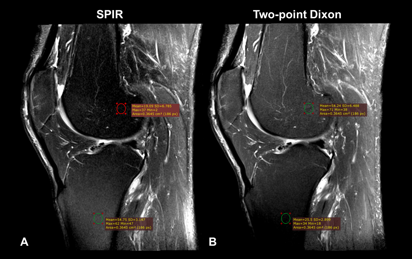
Fat-suppression plays a key role in MR imaging and is used to suppress the hyper-intensity of fat so as to increase contrast between pathological and normal tissue. B1 inhomogeneity affects the uniformity of fat-suppression with “darkness” of fat altered by the B1 variation. For example, fat-suppression in knee MR imaging performed using spectral saturation (such as with the selective partial inversion recovery (SPIR) method1) fat is “lighter” where B1 inhomogeneity results in lower flip-angles (Figure 1A). In contrast, for turbo-spin-echo (TSE) imaging with Dixon2 based fat-suppression, fat is “darker” in regions with lower excitation flip-angles (Figure 1B). Using adiabatic pulses provides robustness to B1 variation with spectral attenuated inversion recovery (SPAIR) fat-saturation3, but the longer duration of the adiabatic RF pulses and inversion time for T1 relaxation increases the scan time with SPAIR. Adiabatic pulses also increase specific-absorption-rate (SAR).
In this work, we propose dual repetition of spectrally selective high bandwidth Shinnar–Le Roux (SLR) RF pulses with different flip-angles to achieve B0 and B1 robust uniform fat-saturation (fat-sat). Multi-pulse saturation methods have been used in other applications such as saturation recovery preparation in cardiac imaging4, but this is the first time to our knowledge that this technique has been used for fat saturation in joint imaging.
THEORY & METHODS
5 subjects were imaged using a single-channel transmit, 15-channel receive knee coil at 3T (Magnetom Skyra, Siemens Healthineers, Erlangen, Germany) using a TSE sequence with typical imaging parameters. Fat-suppression was performed with two-point Dixon, SPIR, SPAIR and Dual flip-Angle (DFA) methods.
Figure 2 shows the schematic for the pulse sequence diagram of the proposed DFA fat-sat method in that the two RF pulses (with flip-angles α1 and α2) were separated by inter-pulse duration and gradient spoilers were placed for the complete dephasing of transverse magnetization between RF pulses.
The two SLR RF pulses had time-duration of 14.8ms each. The DFA fat-sat module had a total duration of 35ms, including the spoilers. In comparison, the SPAIR fat-sat module had duration of ~150ms.
The values of α1 and α2 for the DFA fat-saturation were optimized using Bloch simulations with assumption of instantaneous spin tipping by RF pulse as follows. The residual longitudinal magnetization after the second spoiler was determined for a particular η, the multiplicative factor for flip-angle variation due to B1 inhomogeneity. The difference (Ω) between the ideal (no B1 inhomogeneity) and the achieved transverse magnetization was determined. The value of Ω was integrated for a typical range of η between 0.8 and 1.2 to obtain the cumulative error (Γ). The combinations of α1 and α2 for which the B1 induced error Γ was minimum were determined to be the optimal flip-angle combinations.
RESULTS & DISCUSSION
Figure 3 shows the comparison of fat-sat uniformity between SPIR, SPAIR and the DFA method with flip-angle combinations of 65° and 150°. It can be observed that darkness of fat is different in the tibia compared to the femur for the SPIR method. SPAIR and the dual flip-angle methods provided uniform fat-sat. SPAIR (3min 23 sec) had longer scan time than SPIR (2min 21sec) and the DFA methods (2min 44sec). DFA method provided fat-sat uniformity similar to the SPAIR method with scan time similar to SPIR method.
Spectral saturation based fat-sat methods are affected B0 inhomogeneity. In this work high bandwidth SLR RF pulses were used to improve robustness to B0 variation.
Uniformity of fat-sat is affected by B1 inhomogeneity that is inherent to the combination of imaging RF coil, excitation or saturation pulse, and the position of the imaged object within a coil. Robustness to patient positioning becomes even more important for local transmit coils, which have a smaller B1 homogeneity volume compared to a body coil. Spectral fat-sat methods such as SPIR suffer from B1 variation of the local transmit coils typically used in knee imaging. SPAIR provides uniform fat-sat, but at the cost of increased scan time. The DFA method provided uniform fat-sat with minimal scan time increase (one additional RF pulse plus, gradient spoiling time) by reducing the effect of B1 variation on flip-angles by combining two flip-angles. While two RF pulses are applied, both pulses experience the same B1 inhomogeneity at a given voxel and its effect is cancelled out between the two pulses, producing B1-insensitive longitudinal spin magnetization at time of imaging excitation with optimized two flip angles for a given B1 inhomogeneity range.
CONCLUSION
DFA method provides a novel approach to achieve B1 and B0 robustness in fat-saturation with scan time durations shorter than methods that use adiabatic pulses.Acknowledgements
No acknowledgement found.References
1. OH CH. Selective partial inversion recovery (SPIR) in steady state for selective saturation magnetic resonance imaging (MRI). Abstr Soc. Magn. Reson. Med. San Fr. [Internet] 1988;1042.
2. Dixon WT. Simple proton spectroscopic imaging. Radiology [Internet] 1984;153:189–194. doi: 10.1148/radiology.153.1.6089263.
3. Bernstein M, King K, Zhou X. Handbook of MRI pulse sequences. 2004.
4. Chow et al. Saturation pulse design for quantitative myocardial T1 mapping. JCMR (2015) 17:84. doi: 10.1186/s12968-015-0187-0.
Figures