4008
Separation of Water and Fat during Spin-Lock Magnetic Resonance Imaging with Multifrequency Fat Spectrum Modeling1Department of Imaging and Interventional Radiology, The Chinese University of Hong Kong, Shatin, Hong Kong, 2Department of Diagnostic and Interventional Radiology, Technical University of Munich, Munich, Germany, 3Shenzhen Institutes of Advanced Technology, Shenzhen, China
Synopsis
Spin-lock technologies are useful for probing macromolecular environment of tissue. The conventional spin-lock approaches cannot lock water and fat spins simultaneously due to chemical shift effect of fat, which can lead to artifacts in tissues with fatty infiltration. Based on the latest development of the spin-lock technique, we propose spin-lock Dixon methods to perform water-fat separation in spin-lock MRI. Our methods may offer a solution to perform reliable spin-lock MRI in tissues with fatty infiltration.
Introduction
Spin-lock technologies1 are useful for probing macromolecular environment of tissue. The conventional spin-lock approaches cannot lock water and fat spins simultaneously due to chemical shift effect of fat, which can lead to artifacts in tissues with fatty infiltration. When performing spin-lock, the fat signal is typically suppressed by spectrally selective RF pulses. However, these methods are susceptible to B0 field inhomogeneities. Besides, it cannot completely suppress fat signal since fat has multiple spectral peaks.
Dixon approaches2 for water-fat separation do not suffer from these problems. In this work, we investigate Dixon approaches to perform water-fat separation rather than fat suppression during spin-lock MRI. Our methods may offer a solution to perform reliable spin-lock MRI in tissues with fatty infiltration.
Methods
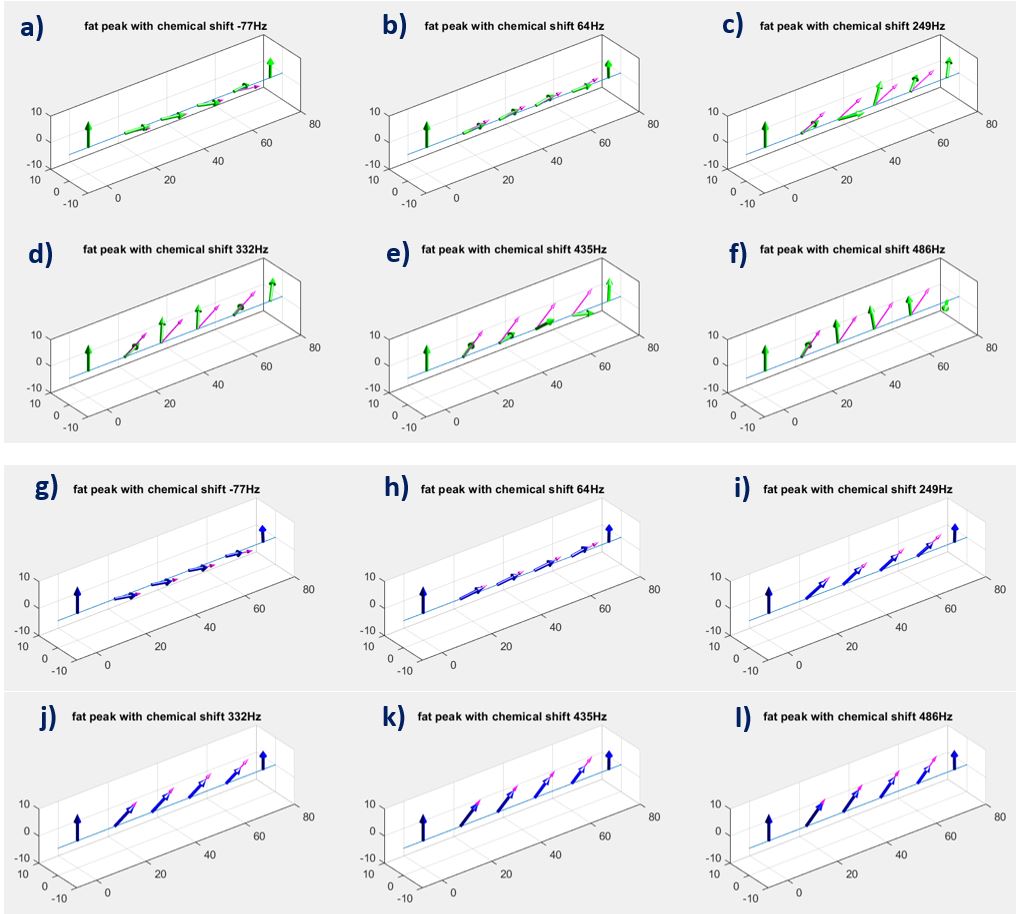
Our design of spin-lock RF pulse cluster is based on the methods reported in3,4. We term this method Adiabatic Continues-wave Constant-amplitude Spin-Lock (ACCSL). We noticed ACCSL can lock water and fat spins simultaneously. We illustrate this in Figure 1 using Bloch simulation.
We investigate two acquisition approaches for Dixon water-fat separation: the conventional spin-lock followed with multiecho acquisition and ACCSL followed with multiecho acquisition. We termed two methods conventional spin-lock Dixon and ACCSL Dixon, respectively.
After spin-lock preparation with a given time-of-spinlock (TSL), the images acquired at echo time TE can be expressed as:
s(r,TE,TSL)=(ρw(r,TSL) + ρf(r,TSL)Σn [βn(TSL) ej2πfnTE])ej2πψ(r)TE,
where ρw and ρf are water and fat image, respectively; ψ(r) is the field map; fn is the chemical shift of the nth fat peak; and βn(TSL) is the relative amplitude of the nth fat peak. βn(TSL) is normalized such that Σnβn(TSL) = 1. T2* decay can be included in the equation using the method described in5.
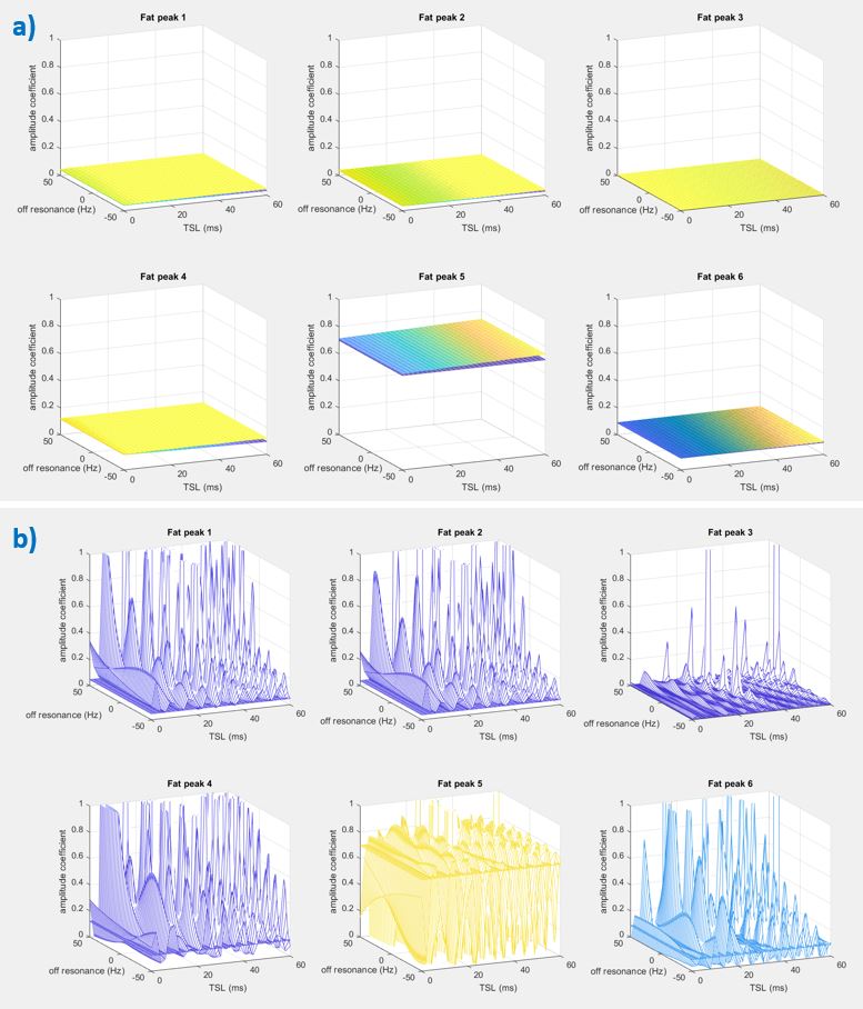
Note βn(TSL) depends on the TSL due to the difference of magnetization evolution of each individual fat peak during the spin-lock. Figure 2 shows full equation Bloch simulation of βn(TSL) using the conventional spin-lock and the ACCSL. Note for the conventional spin-lock, the failure of spin-lock of fat peaks results in significant oscillation, which leads to unpredictable amplitude coefficients βn. When using ACCSL, the magnetization of fat peaks follows regular exponential relaxation4. After normalization, the relaxation effect during TSL is mostly removed, which results in relatively flat βn. Thus, with ACCSL Dixon acquisition, under the first order approximation, we can use TSL-independent amplitude coefficients of a multi-peak fat model to perform the water-fat separation. For circumstances that this approximation is violated, a pre-calibration procedure6,7 can be performed to obtain βn.
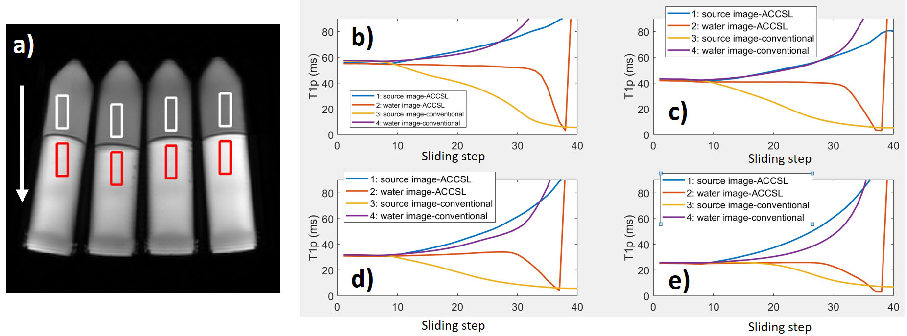
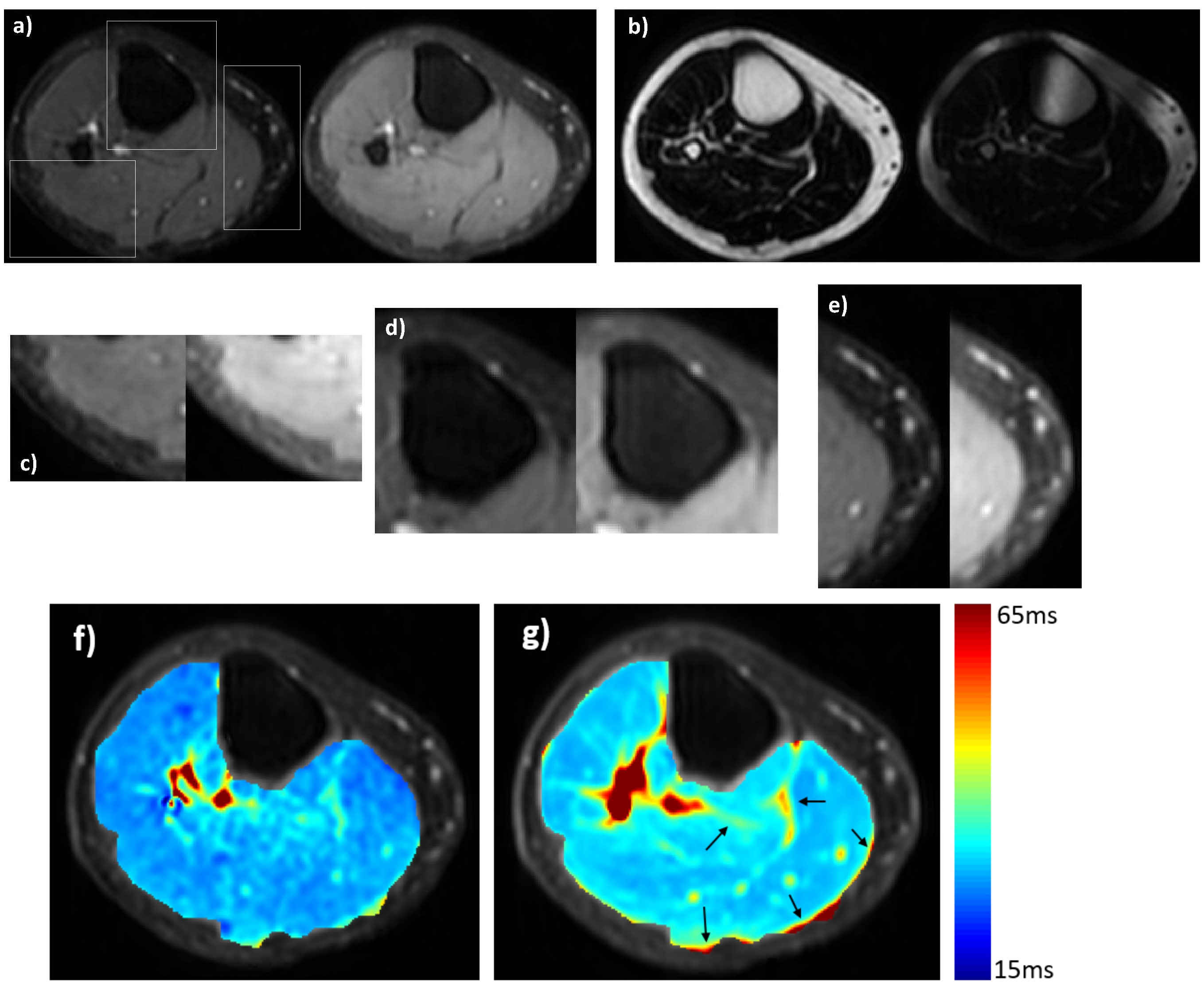
Simulation data were generated using Bloch equations to demonstrate the proposed spin-lock Dixon methods. Phantom and in vivo calf muscle scan were conducted on a Philips Achieva TX 3.0T scanner (Philips Healthcare, Best, the Netherlands). An 8-channel head coil and an 8-channel T/R knee coil (Invivo Corp, Gainesville, USA) were used for phantom and in vivo scan, respectively. Volunteer scan was conducted under the approval of the Institutional Review. For phantom study, we choose an ROI and slide it across the water-fat interface to create various levels of the fat fraction within the ROI (Figure 4). T1rho value is then calculated from these ROIs. A 2D multi-shot Fast Spin Echo sequence8 was used for data acquisition. Four TSL 0, 10, 20, 50ms, spin-lock frequency 500Hz, and 3-point Dixon acquisition with delta TE 0, 1, 2ms were used for simulation, phantom, and in vivo studies. Other imaging parameters included TR/TE 2000/7.5ms, resolution 0.65mm x 0.65mm, slice thickness 5 mm, and echo train length 10. The Dixon reconstruction was performed using the method reported in6. The relaxation model described in4 and the standard mono-exponential model was used for T1rho quantification with ACCSL Dixon acquisition, and the conventional spin-lock Dixon acquisition, respectively.
Results and Discussion
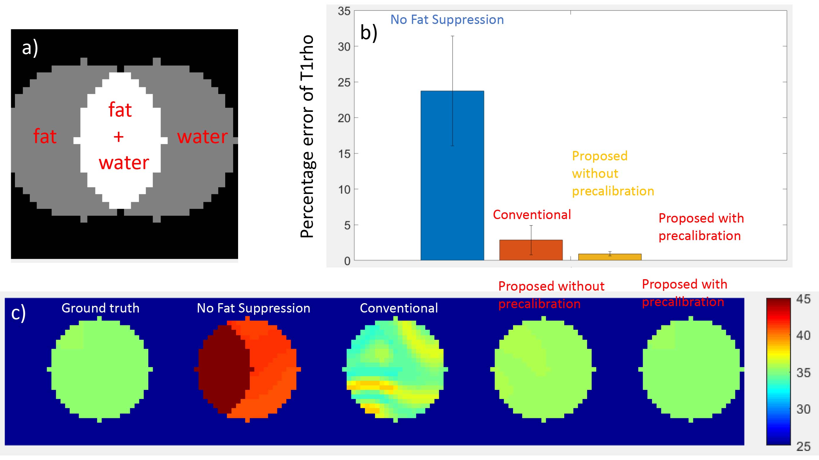
Figure 3, 4, and 5 show simulation, phantom, and in vivo results, respectively. In the simulation study, conventional spin-lock Dixon significantly reduces T1rho quantification error compared to T1rho obtained using the source images (no fat suppression). ACCSL Dixon without precalibration further reduces the error. With precalibration, ACCSL Dixon achieves nearly zero error in this simulation study.
The results from phantom scanning are consistent with simulation results. ACCSL Dixon shows greater robustness compared to conventional spin-lock Dixon. T1rho value calculated from source images is unreliable when ROI has mixed water and fat.
For in vivo results, ACCSL Dixon shows more accurate water-fat separation compared to conventional spin-lock Dixon. T1rho quantification using ACCSL Dixon is less susceptible to fat infiltration compared to the conventional spin-lock Dixon.
Conclusion
We reported spin-lock Dixon approaches to perform water-fat separation during spin-lock MRI. It has the potential to address the problems associated with the conventional fat suppression methods used in spin-lock MRI.Acknowledgements
This study is supported by a grant from the Innovation and Technology Commission of the government of Hong Kong SAR (Project ITS/051/17) and a grant from the Research Grants Council of the Hong Kong SAR (Project SEG CUHK02). We would like to acknowledge general support from Philips Healthcare.References
1. Sepponen RE, Pohjonen JA, Sipponen JT, Tanttu JI. A method for TIp imaging. J Comput Assist Tomogr. 1985;9(6):1007‐1011. https://doi.org/10.1097/00004728‐198511000‐00002
2. Dixon WT. Simple proton spectroscopic imaging. Radiology 1984;153:189–194.
3. Chen W. Artifacts correction for T1rho imaging with constant amplitude spin-lock. Journal of Magnetic Resonance. 2017 Jan 1;274:13-23.
4. Jiang B, Chen W. On‐resonance and off‐resonance continuous wave constant amplitude spin‐lock and T1ρ quantification in the presence of B1 and B0 inhomogeneities. NMR in Biomedicine. 2018 Apr 25:e3928.
5. Yu H, McKenzie CA, Shimakawa A, Vu AT, Brau AC, Beatty PJ, Pineda AR, Brittain JH, Reeder SB. Multiecho reconstruction for simultaneous water‐fat decomposition and T2* estimation. Journal of Magnetic Resonance Imaging. 2007 Oct 1;26(4):1153-61.
6. Yu H, Shimakawa A, McKenzie CA, Brodsky E, Brittain JH, Reeder SB. Multiecho water‐fat separation and simultaneous R estimation with multifrequency fat spectrum modeling. Magnetic Resonance in Medicine. 2008 Nov;60(5):1122-34.
7. Weidlich D, Diefenbach MN, Schlaeger S, Hock A, Ruschke S, Karampinos DC. In-vivo water T2 mapping in tissues containing water and fat using a T2-prepared 3D Dixon TSE sequence and a pre-calibrated at spectrum model. ISMRM 27th annual meeting, 2018, Paris, France, 4223
8. Chen W, Chan Q, Wáng YX. Breath-hold black blood quantitative T1rho imaging of liver using single shot fast spin echo acquisition. Quantitative imaging in medicine and surgery. 2016 Apr;6(2):168.
Figures