4003
Microfluidics preparation of liposome containing alginate hydrogel microbeads for CEST MRI1City University of Hong Kong, Hong Kong, China
Synopsis
Image-guided cell therapy plays an important role in monitoring and adjusting the treatment regimen. We have demonstrated that cell viability could be monitored by CEST pH-nanosensors in alginate hydrogel microbeads. Here, we investigated the use of microfluidics to generate liposome containing microbeads and examined their CEST properties after loading with the iodine CT contrast agents Iohexol and Iopamiro. We have successfully demonstrated that the microbead size has greatly reduced from 300-400 μm to 20 μm. Moreover, iodine agents loaded liposomal microbeads generated 4% CEST contrast at 4.2 ppm at 3T. These findings demonstrate a robust platform for the fabrication of liposome containing microbeads under CEST MR guidance.
Introduction
Cell encapsulation using hydrogel has emerged as a promising treatment for many diseases and disorders. Microfluidics can fabricate highly monodispersed microbeads at a wide range from several microns to hundreds of microns in diameter. It can be tailored to match all the major gelations and can be designed to genearate a variety of microstructurs.[i],[ii] We have demonstrated that the design of microbeads could facilitate the monitoring of cell viability using CEST MRI. It detects the decrease in CEST contrast when cell death occurred.[iii],[iv] In this study, we aim to fabricate microbeads with smaller size and slow down the release of contrast agents from liposomes[v]. We developed a robust microfluidic platform for the fabrication of liposome-contained alginate microbeads, which iodine CT contrast agents were loaded into the liposomes, and studied their CEST contrast at 3T. The results showed that this robust microfluidic platform generated microbeads at a few tens of microns, which could faciliate the monitoring cell status after cell transplantation.Methods
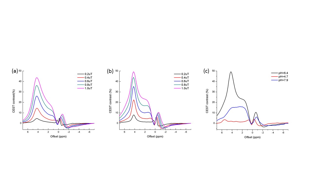
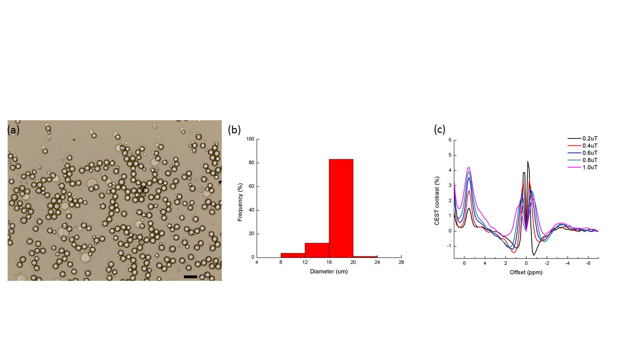
Liposomes were prepared by using thin film hydration method.[i] In brief, EPC, cholesterol and DSPE-PEG2000 were mixed in a molar ratio of 65:32:3 in chloroform with total weight of lipids of 25mg. The solvent was removed and the resulting thin film was hydrated with 1 mL Iohexol solution or 1mL Iopamiro solution.The suspension was annealed and extruded. Liposome was mixed with 4wt% alginate solution and 200mM Ca-EDTA solution at volume ratio of 2: 1: 1, as the water phase while mineral oil with 2wt% span 80 was the oil phase. Microfluidic devices were fabricated by soft lithography and replica modeling of PDMS.[ii] After microbead collection, acetic acid was added to form hydrogel beads.[iii] Phantoms were imaged on a horizontal bore 3T preclinical Bruker BioSpec system at 37 oC. Image was acquired using RARE with the following saturation parameters: B1 were 0.2, 0.4, 0.6, 0.8 and 1.0uT and Tsat=3000ms; and imaging parameters: Slice thickness=2 mm, RARE factor = 32, repetition time/echo time (TR/TE) = 6000/4.7 ms, -10 to + 10 ppm, 0.2 ppm steps. CEST contrast was calculated by applying Lorentzian fitting on Z spectra (CESTcontrast%). [i] Chan, K. W. et al. J Control Release 2014:180:51-59. [ii] Y. Xia, et al, Angewandte Chemie International Edition, 1998, 37, 550-575. [iii] Choi CH, et al, Biomedical Microdevices. 2007;9:855-62. Figure 1. CEST contrast of Iohexol liposome (a) and Iopamiro liposomes (b) and pH dependency of Iopamiro liposome (c) at 3T. Figure 2. Alginate beads prepared with microfluidic device (scale bar=40μ m) (a) and size distribution of alginate beads prepared by oil and water flow rate of 700μl/h and 150μl/h, respectively (b), and CEST contrast of alginate hydrogel microbeads containing Iopamiro-liposome at pH 4.1 at 3T (c).Results
The prepared Iohexol-liposomes and Iopamiro-liposome showed CEST contrast of 44% and 49% at 4.2ppm and B1 = 1.0 μT, respectively. Their particle size and polydispersity index were 146 nm, PDI=0.150 and 135nm, PDI= 0.107 respectively. The particle concentration was at (2.7±0.3)x1018 for the CEST measurement before transferring them to incorporate into the alginate microbeads by microfluidics. The alginate beads prepared by the designed microfluidics device at oil flow rate of 700 μl/h and water flow rate of 150 μl/h had size of 17.4 μm with CV of 11.3%. The LipoCEST microbeads containing Iopamiro-liposomes show CEST contrast of 4% at 5.6ppm.Discussion
The liposome containing alginate microbeads prepared by microfluidics had a much smaller size (20μm) and monodisperse as compared to those prepared by electrospray approach4. The prepared Iohexol-liposomes and Iopamiro-liposome showed strong CEST contrast almost 50% contrast at 4.2ppm, which is favorable for the preparation of alginate microbeads. The liposomal alginate beads containing the iodine CT CAs as prepared by our microfuildics platform showed distinctive CEST contrast, which could be valuable for monitoring status after implantation. The further optimizations of the platform to generate liposomal microbeads with higher CEST contrast are underway.Conclusion
We have developed a robust platform to fabricate liposome-contained alginate microbeads with small size and narrow size distribution. By incorporating the iodinate CT contrast agents, i.e. Iohexol and Iopamiro, we showed that these liposomal microbeads generated unique CEST contrast at 4.2 ppm. We believe this platform can facilitate MR guided cell therapy, especially to improve the injectability and detectability of the CEST microbeds.Acknowledgements
This study was supported by CityU: P9610362; P7200516; P6000612;P7004859; RGC: GRF-9042620; NSFC: 81871409-H1808.References
[1] Teh SY, et al. Lab on a Chip. 2008;8:198-220.
[2] Shintaku H, et al. Microsystem Technologies. 2006;13:951-8.
[3] P. C. Van Zijl, et al, Magnetic Resonance in Medicine, 2011, 65, 927-948.
[4] K. W. Chan, et al , Nature Materials, 2013, 12, 268-275.
[5] Kim J, et al. Angewandte Chemie. 2011;50:2317-21.
[6] Chan, K. W. et al. J Control Release 2014:180:51-59.
[7] Y. Xia, et al, Angewandte Chemie International Edition, 1998, 37, 550-575.
[8] Choi CH, et al, Biomedical Microdevices. 2007;9:855-62.
Figures