3986
GlucoCEST MRI detects metabolic degradation in the mouse brain after rapid microwave fixation1Howard University, Washington, DC, United States, 2Children's National Medical Center, Washington, DC, United States, 3Fu Jen Catholic University, New Taipei City, Taiwan, 4Yuan Ze University, Taoyuan City, Taiwan
Synopsis
The study of neurometabolites and the detection of glucose in the brain has relied on anesthetized animals. Prolonged periods of anesthesia required for these studies may affect these metabolites and the ability to interpret glucose detection in the brain. However, performing post-mortem studies of neurometabolites and glucose detection in the brain is difficult to interpret because of rapid degradation. In this study, we utilized focal beam microwave irradiation (FBMI) without anesthesia to collect brain tissue and determine whether there is postmortem degradation of neurometabolites and glucose using 1H-NMR and glucoCEST.
Introduction
The study of neurometabolites and the detection of glucose in the brain has relied on anesthetized animals. Prolonged periods of anesthesia required for these studies may affect these metabolites and the ability to interpret glucose detection in the brain [1]. However, performing post-mortem studies of neurometabolites and glucose detection in the brain is difficult to interpret because of rapid degradation [2]. In this study, we utilized focal beam microwave irradiation (FBMI) without anesthesia to collect brain tissue and determine whether there is postmortem degradation of neurometabolites and glucose using 1H-NMR and glucoCEST.Materials and Methods
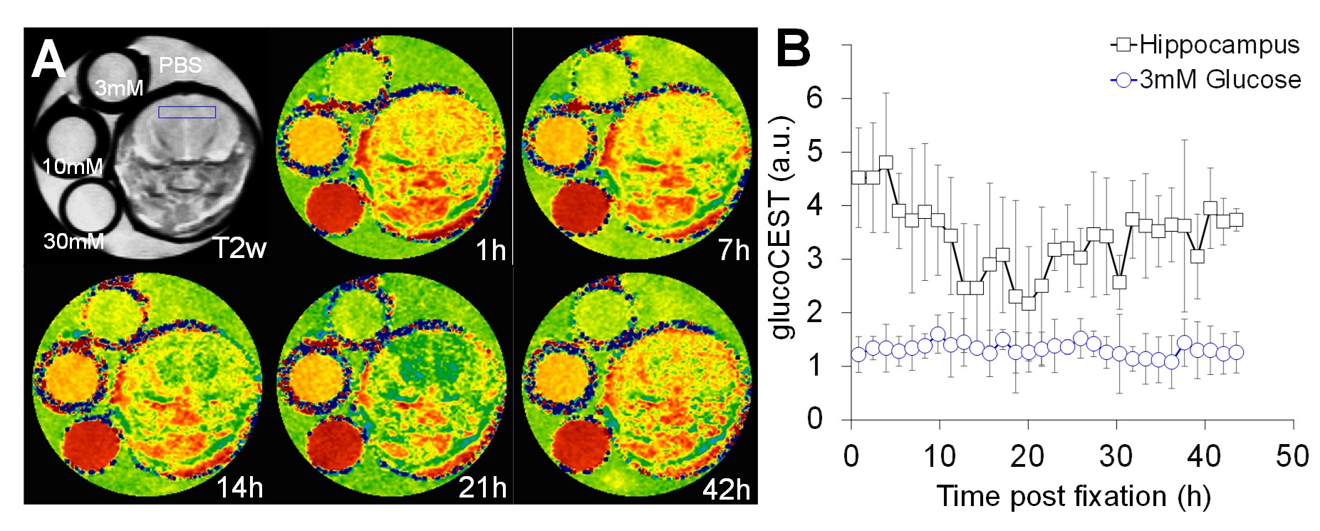
Five C57/BL6 mice were administered 0.55 mg/g of D-glucose through intraperitoneal injection in the awake state 15 minutes prior to sacrifice using FBMI. No anesthetic agents were utilized. The head was quickly decapitated and the brain was kept in the skull. The tissues were immediately transferred for 1H-NMR and glucoCEST scans using a Bruker 9.4T spectrometer. The tissue was wrapped with Parafilm and immersed in PBS solution for imaging with three glucose phantoms in 3mM, 10mM, and 20mM concentration over 48 hours after fixation with FBMI. The localized 1H-NMR were acquired using a PRESS sequence for hippocampus. The NMR data were analyzed using LCModel for extracting NAA, Lac, Cho, Myo-Inositol (Ins), Taurine (Tau), and Glutamate-Glutamine (Glx) levels. The glucoCEST data were acquired by fast spin-echo sequence with MT pulse module (2µT, 2000ms). The MT offset frequences (Δω) were set from -1.6kHz to +1.6kHz with 40Hz stepping to sample 81 points covering the frequency offset range from -4ppm to +4ppm to detect the proton metabolites of glucose. CEST data were calibrated for B0/B1 field inhomogeneity using WASSR [3] technique (0.35µT, 700ms) and spectral interpolation. The glucoCEST contrast were derived by asymmetry of magnetization transfer ratio (MTRasym) calculating the area under the curve at 1.2ppm, 2.1ppm, 2.9ppm for mapping the glucose levels in brain overtime [4].Results
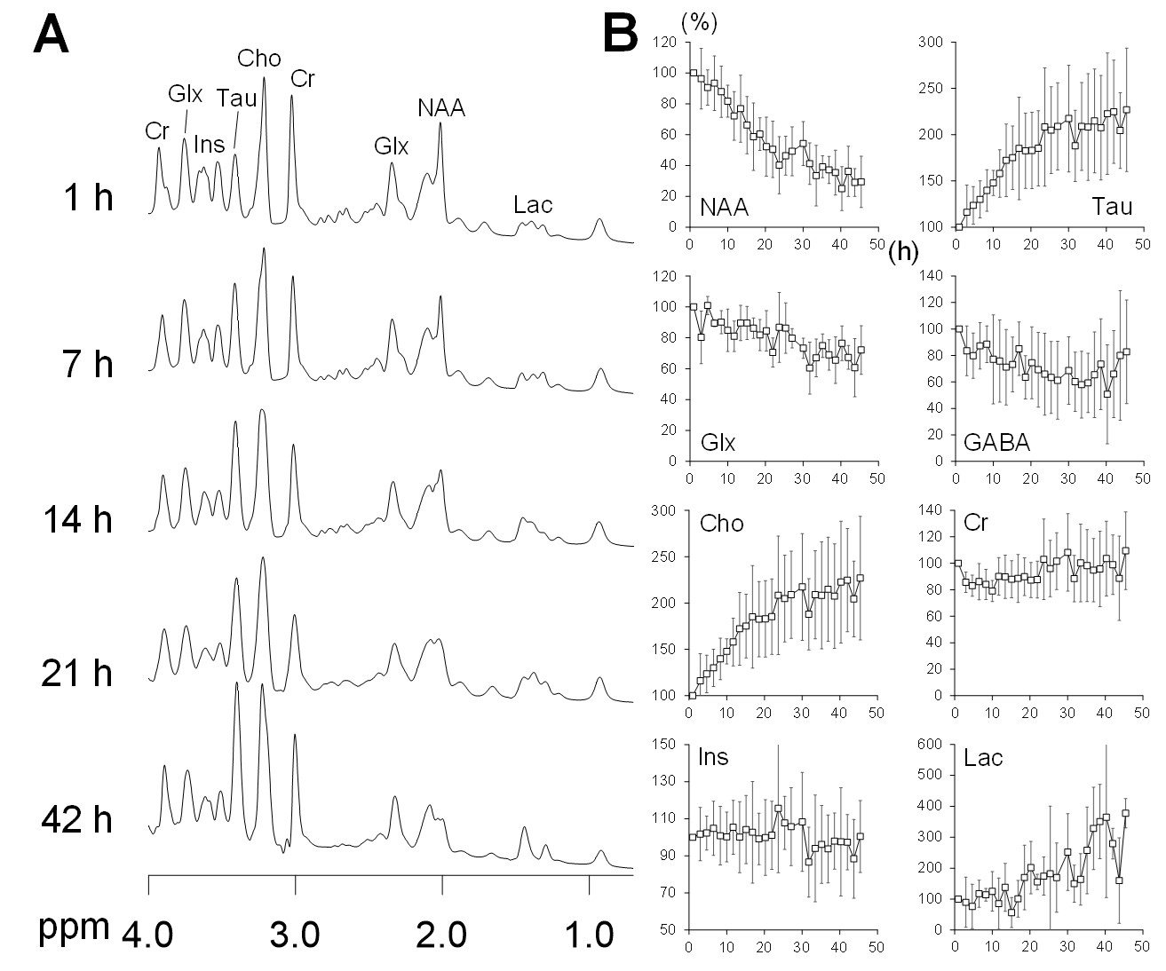
1H-NMR detected a steep decreasing trend of NAA level in brain within 24 h after fixation (Fig.1). The decreasing trend was slower from 24 h to 48 h, while Tau, Cho, and Lac consistently increased over time. Glx showed a decreased trend within 48 h of time and Ins was not different after microwave fixation. The glucoCEST contrast in the 3mM glucose phantom stably maintained throughout the entire experiments (Fig. 2). The glucoCEST detected a decreasing trend of glucose levels in hippocampus within 24 h and a slightly increasing trend from 24 h to 48 h.Discussion and Conclusion
To perform any metabolic functional studies, it is pertinent to collect tissue without concerns for metabolic degradation of signal or influence of anesthetic agents, all of which adversely affect mitochondria. This study shows the feasibility of applying glucoCEST following microwave fixation to detect the glucose level in the brain, which is usually unobservable by 1H-NMR. The decrease of NAA indicated the degradation of neuronal cells, while the increase of Tau, Cho, and Lac reflected the accumulation of metabolic products in anaerobic glycolysis after death. The glucoCEST complimented the 1H-NMR to identify the post-mortem changes of glucose level associated with NAA degradation and Lac production in anaerobic glycolysis during tissue degradation.Acknowledgements
No acknowledgement found.References
[1] Choi IY, et al. J Neurochem 2004;91,778-787;
[2] de Graaf R, et al. J Neurochem 2009;109,494-501;
[3] Kim M, et al. Magn Reson Med 2009;61:1441-1450.;
[4] Tu et al., Sci.Rep. 2018; 8:669
Figures