3978
Labyrinthine lymph MR imaging of patients with vertigo disease: an imaging comparison study with intratympanic or intravenous gadolinium injection1Department of Radiology, Shandong Provincial Hospital Affiliated to Shandong University, Shandong, China, 2GE Healthcare, MR Research China, Beijing, China, 3Gong Ruozhen Innovation Studio, Shandong Medical Imaging Research Institute Affiliated to Shandong University, Shandong, China, 4Shandong Provincial Hospital Affiliated to Shandong University, Shandong, China
Synopsis
In this study, we aimed to determine whether intratympanic (IT) or intravenous (IV) injection of gadolinium is more suitable for labyrinthine lymph imaging of patients with vertigo disease. The corresponding MR images with IV and IT injection were thus systematically compared. While comparable detection of endolymphatic hydrops were shown using both injection methods, higher image contrast between affected and unaffected sides of perilymph region were found using IV method than using IT method. We therefore, demonstrated that IV method can be further applied in clinical diagnosis for patients with vertigo disease.
Introduction
Contrast enhanced MRI has been applied to visualize labyrinthine lymph for patients with vertigo disease1-4. For strong image contrast, an intratympanic (IT) method is usually used for contrast agent injection1-3, although this was known as an invasive examination. To avoid this issue, an intravenous (IV) method, as a non-invasive alternative, has been recently reported for contrast injection in labyrinthine lymph imaging 4.
Based on the advantages of each method, it is, however, still unknown that which one is better for labyrinthine lymph imaging. To determine this, the main aim of this study was, therefore, to compare the respective labyrinthine lymph imaging with IT and IV injection methods systematically, including the signal enhancement in perilymph region and the detection of endolymphatic hydrops (EH).
Materials and methods
Subjects
In total thirty-six patients with unilateral vertigo disease were recruited in this study. Eighteen (49±14 years-old) of them were randomly selected and underwent a bilateral IT injection of gadolinium chelate. The applied agent was diluted 8-fold with saline and injected 24hrs prior to the MR experiments1. The rest eighteen patients (50±15 years-old) were then injected with the gadobenate dimeglumine (0.2mL/kg body weight) using an IV-method 4hrs before the MR experiments5.
MR experiments
All experiments were performed on a 3T clinical scanner (Discovery 750w, GE Healthcare, USA) equipped with an 8-channel phased-array coil. A 3-dimensional CUBE-FLAIR (fluid-attenuated-inversion-recovery) sequence was employed for labyrinthine lymph imaging with scan parameters of repetition time=9000ms; echo time=82.3ms; inversion time=2500ms; echo train length=140; bandwidth=71Khz; matrix size=256×256; NEX=2; slice thickness=1.6mm, and field of view=21x16cm. The scan time was 5 minutes 46 s.
Data analysis
All data were processed at a workstation (Advantage workstation 4.6; GE Medical Systems). For all patients with either IV or IT injection, the MR signal intensities in the bilateral perilymph regions (i..e, with and without affection) were, respectively, evaluated using a defined CM ratio1,6:
CM ratio = SIcochlea/ SImedulla oblongata (Eq.1)
, where SIcochlea represents the image intensity of the affected or unaffected side of perilymph, and SImedullar oblongata is the image intensity of the medullar oblongata served as a reference due to stable MR signal.
In addition, the number of EH in labyrinthine, including in cochlea, in vestibule or in both regions together , were separately determined for each patient. The corresponding ratios of EH number (EHN) to patient number were then calculated.SPSS 20.0 (IBM, Chicago, IL) was used for statistical analyses. The embedded paired-t-test toolbox was applied to compare the CM ratios between the affected and unaffected sides of perilymph region for both IV and IT injection groups, respectively. Also, these regions-specific CM ratios were compared correspondingly between IV and IT injection groups using a paired-test. In addition, a chi-squared test was used to evaluate the EHN ratios between the two injection groups.
For all analysis, the significant threshold was set as P=0.05.
Results
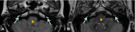
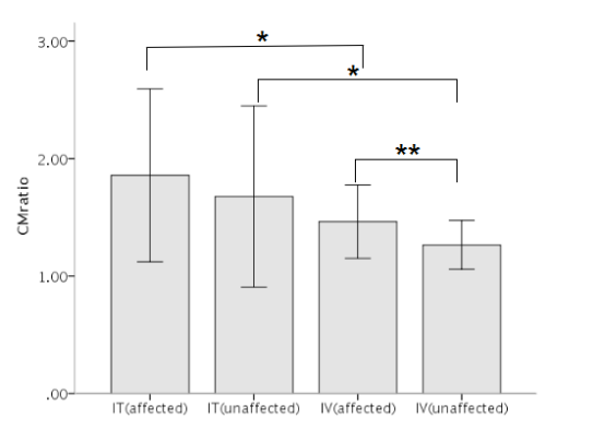
For the IT injection group, comparable CM ratios were found between the affected and unaffected sides of perilymph regions (1.86 ± 0.74 vs 1.68 ± 0.77, P=0.805, Figs 1,2). For the patients injected with IV method, a significant difference for CM ratios was, however, observed between the affected and unaffected sides of perilymph regions (1.46 ± 0.31 vs 1.26 ± 0.21, P=0.001, Figs.1,2).
Meanwhile, comparing the region specific CM ratios between IT and IV groups, significantly higher values were shown in both the affected (1.86 ± 0.74 vs 1.46 ± 0.31, P=0.044) and the unaffected (1.68 ± 0.77 vs 1.26 ± 0.21, P=0.032) sides of perilymph for IT group than for IV group (Figs. 1,2).
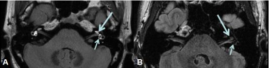
In addition, comparable cochlear EHN ratios (0/18 vs 0/18), vestibular EHN ratios (5/18 vs 7/18) and EHN ratios in both regions together (13/18 vs 11/18, P=0.480, Fig.3) were, respectively, observed between IT and IV injection groups.
Discussion and conclusion
In this study, both intratympanical and intravenous methods for contrast agent injection were investigated in labyrinthine imaging for patients with unilateral vertigo disease. Using IV method, significantly higher image contrast was found in the affected than unaffected sides of perilymph regions, while comparable image intensities were shown between the affected and unaffected sides for IT method. This discrepancy might be due to more effected permeability of the blood-labyrinth barrier using IV method than that of the round window by IT method7. In addition, comparable detection for endolymphatic hydrops in cochlea and vestibule was observed using both methods.
In conclusion, due to high image contrast between the affected and unaffected sides of perilymph and comparable detection of EH , IV method, compared to IT injection, can thus be suggested to be further applied in clinical diagnosis for patients with vertigo disease.
Acknowledgements
No acknowledgement found.References
[1] Nakashima T, Naganawa S, Sugiura M, et al.Visualization of endolymphatichydrops in patients with Me´nie`re’s disease.Laryngoscope.2007;117:415–420.
[2] Naganawa S, Sugiura M, Kawamura M, et al.Imaging of endolymphatic andperilymphatic fluid at 3T after intratympanic administration of gadoliniumdiethylene-triamine pentaacetic acid.AJNR Am J Neuroradiol.2008;29:724–726.
[3] Naganawa S, Satake H, Iwano S, et al.Imaging endolymphatic hydrops at 3Tesla using 3D-FLAIR with intratympanic Gd-DTPA administration.MagnReson Med Sci.2008;7:85–91.
[4] Naganawa S, Nakashima T. Cutting edge of inner ear MRI.Acta Otolaryngol Suppl 2009;560:15–21.
[5] Naganawa S, Komada T, Fukatsu H, Ishigaki T, Takizawa O. Observation of contrast enhancement in the cochlearfluid space of healthy subjects using a 3D-FLAIR sequence at 3 Tesla. Eur Radiol 2006;16:733–737.
[6] Yamazaki M, Naganawa S, Kawai H, et al.Signal alteration of the cochlear perilymph on 3 different sequences after intratympanic Gd-DTPA administration at 3 Tesla: comparison of 3D-FLAIR, 3D-T1-weighted imaging, and 3D-CISS.Magn Reson Med Sci2010;9:65–71.
[7] Tagaya M, Yamazaki M, Teranishi M, et al.Endolymphatic hydrops and bloodlabyrinth barrier in Me ´nie`re’s disease.Acta Otolaryngol.2011;131:474–479.
Figures