3977
Magneto-Caloric Materials as Symmetric Reverse-Contrast Switchable MRI Labels1Applied Physics and Instrumentation Group, HHMI-Janelia Reseach Campus, Ashburn, VA, United States, 2Electrical and Computer Engineering Department, United States Naval Academy, Annapolis, MD, United States, 3Laboratory of Functional and Molecular Imaging, NIH/NINDS, Bethesda, MD, United States, 4NIH Mouse Imaging Facility, NIH/NINDS, Bethesda, MD, United States
Synopsis
We describe the use of magneto-caloric materials as symmetric reverse-contrast switchable MRI labels at physiological temperatures. We present physical and temperature tunable MRI measurements on two different magneto-caloric materials, Iron-Rhodium (Fe-Rh) and Lanthanum-Iron-Silicon (La-Fe-Si) that both have sharp first-order magnetic phase transitions at the same bias DC magnetic field of 1 Tesla and physiological temperature of 37°C (310K), but with simultaneously positive and negative slope of magnetization vs. temperature, respectively, and therefore reverse image contrast in MRI. Thus, we show that different magneto-caloric materials provide an opportunity for the development of versatile multi-functional high differential contrast ratio switchable MRI labels.
Introduction
Development of novel contrast mechanisms and labeling agents for MRI is critical for further advancements in non-invasive cell imaging, tracking, and readout of physiological conditions in-vivo1-5. We have recently proposed6 that magneto-caloric materials (developed for magnetic refrigeration7-9, data storage10, and spintronics applications11) may provide an opportunity for development of tunable and switchable high-contrast labels for MRI. We showed in the proof-of-concept experiments that the extremely sharp first-order magnetic phase transitions these materials have at physiological temperatures and in the presence of the large Tesla-scale DC magnetic field values associated with MRI scanners provide an ideal match to the requirements for the design of novel high-contrast ratio switchable MRI labels12. Here we further exploit the properties of magneto-caloric materials to demonstrate symmetric reverse-contrast switchable MRI labels. We present physical and MRI measurements on two different classes of magneto-caloric materials, Iron-Rhodium (Fe-Rh)13,14 and Lanthanum-Iron-Silicon (La-Fe-Si)15,16. They both have sharp first-order magnetic phase transitions at the same bias DC magnetic field of 1 Tesla and physiological temperature of 37°C (310K), but with simultaneously positive or negative slope of magnetization vs. temperature, respectively.Methods
Iron-Rhodium granules (Fe 49%, Rh 51% atomic composition, 99% purity) were prepared by mixing in an arc melting furnace (American Elements Corp.), followed by high-temperature annealing in Argon gas furnace at 1,000°C for two weeks, and subsequently quenched in ice-water. The samples were then cut into mm-scale discs and polished. Lanthanum-Iron-Silicon (La-Fe-Si) powder sample (100μm-250μm particle size) was obtained from a commercial vendor (Calorivac H product line from Vacuumschmelze). Temperature dependent magnetic properties of both samples were measured in a vibrating sample magnetometer (Versalab System from Quantum Design, Inc.). A 1T benchtop MRI (ICON system) was used for MRI characterization of the samples as this was the polarizing DC magnetic field where the sharp first order transition occurs near the physiological temperature of 37°C (310K) for both material systems. For the MRI characterization, the samples were mounted side by side on a glass slide which was placed inside a 15mL centrifuge tube next to a MRI-compatible optical fiber-based thermometer (FISO Technologies, Inc.). The sample tube was wrapped in water tubing connected to a temperature controlled water circulating bath in order to sweep and control the temperature of the samples and their environment around physiologically relevant conditions (25°C-55°C).Results
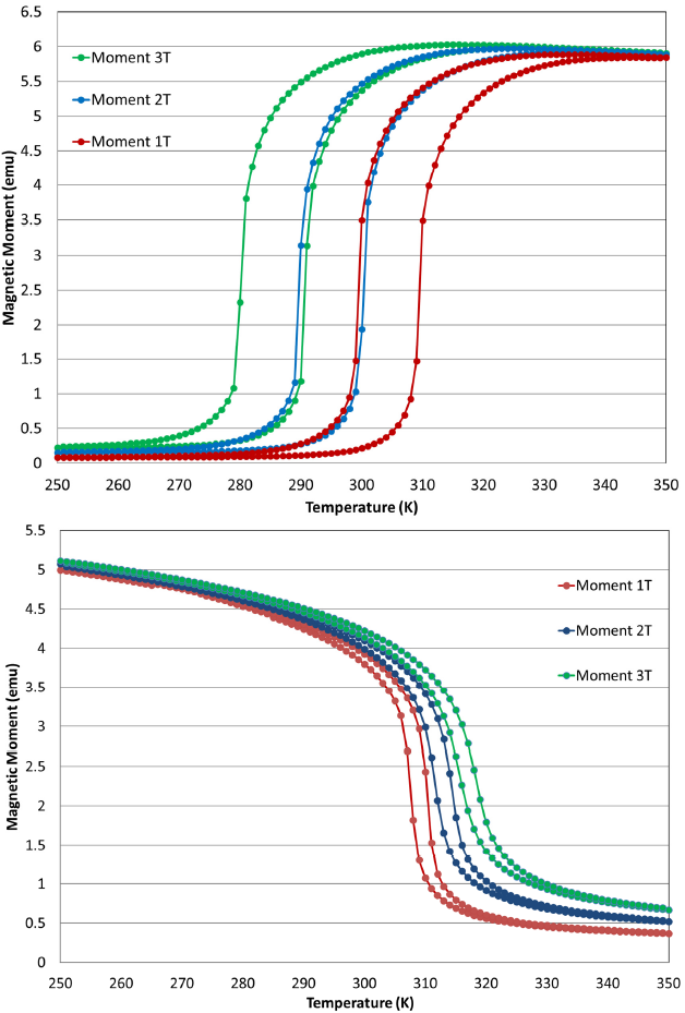
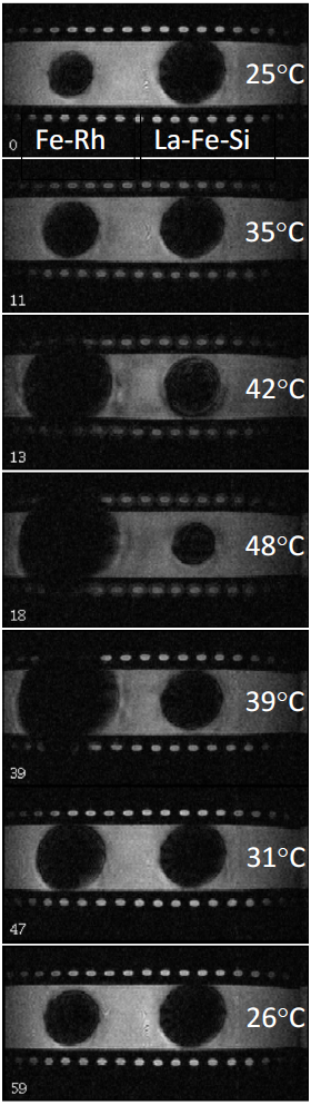
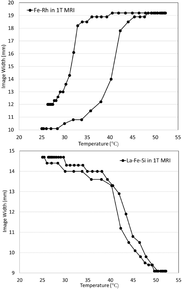
Figure 1(a) shows the vibrating sample magnetometer measurements of the magnetic moment of the 99% purity Fe-Rh disk sample as a function of temperature in different bias DC magnetic fields. The sample exhibits a sharp transition from an antiferromagnetic to a ferromagnetic state over a very narrow range of physiologically relevant temperatures. More specifically, the sample has a sharp positive slope transition around physiological temperature (37°C = 310K) in the field of around 1 Tesla (red curve). Figure 1(b) shows the measurements of the magnetic moment of the La-Fe-Si sample as a function of temperature in the same set of DC magnetic fields. This sample also exhibits a sharp transition over a narrow range of physiologically relevant temperatures, but this time with a narrower hysteresis and a steep negative slope of M vs. T at the physiological temperature (37°C = 310K) and field of 1 Tesla (red curve). Figure 2 shows a sequence of representative gradient-echo images of the effect of the samples on the surrounding Mn-doped water as the entire phantom is heated from 25°C to 53°C and then cooled. The opposite sense of the MRI contrast changes for the two materials due to the opposite slope of their moment vs. temperature properties is clearly evident and closely follows the magnetic properties shown in Figure 1(a) and 1(b). (Image parameters: TR/TE = 39/1.7 ms, FA = 30 degrees, nominal resolution = 0.35 x 0.35 x 0.8mm, FOV = 67.2 x 33.6 x 12.8 mm, Acquisition BW = 125000 kHz, B0 direction is through plane). Figure 3 shows the measurement of the width of the image of each magneto-caloric sample created by the signal loss due to the magnetic field gradients from the Fe-Rh and La-Fe-Si samples as temperature is varied.Discussion and Conclusion
Symmetric but opposite simultaneous image contrast changes in two magneto-caloric material systems of Fe-Rh and La-Fe-Si at the same 1 Tesla DC magnetic field at physiological temperatures were clearly demonstrated. Such demonstrations provide further impetus for the development of versatile multi-functional high differential contrast ratio switchable MRI labels using magneto-caloric materials. Developments in preparing such materials in microparticle form, tuning their physical properties through alloying, and designing the temperature and magnetic field instrumentation for controlled switching of these materials for in-vivo use will be discussed.Acknowledgements
This research was supported by the Howard Hughes Medical Institute, United States Naval Academy, and the NINDS Intramural Research Program of the National Institutes of Health. We thank Tim Harris of HHMI for initial support of this work, Neil Dilley of Quantum Design company for initial magnetometry assistance, Barbara Marcheschi and Alan Huston of the Naval Research Laboratory for initial sample preparation help, and Brian Bell and Kevin Ackerman of Vacuumschmelze company for generously providing us with the Calorivac H samples of La-Fe-Si. We thank Murali Cherukeri of NIH for use of the 1T MRI system.References
1. E. M. Shapiro, S. Skrtic, K. Sharer, J. M. Hill, C. E. Dunbar, and A. P. Koretsky, MRI detection of single particles for cellular imaging, Proc. Natl. Acad. Sci. U S A. 101, 10901 (2004).
2. J. W. M. Bulte, D. L. Kraitchman, Iron oxide MR contrast agents for molecular and cellular imaging. NMR Biomed. 17,484 (2004).
3. G. Zabow, S. Dodd, J. Moreland, and A. P. Koretsky, Micro-engineered local field control for high-sensitivity multispectral MRI, Nature 453, 1058 (2008).
4. E. T. Ahrens and J. W. M. Bulte, Tracking immune cells in vivo using magnetic resonance imaging, Nature Reviews Immunology 13, 755 (2013).
5. G. Zabow, S. Dodd, and A. P. Koretsky, Shape-changing magnetic assemblies as high-sensitivity NMR-readable nanoprobes, Nature 520, 73 (2015).
6. M. Barbic, T. D. Harris, S. Dodd, H. D. Morris, A. P. Koretsky, B. Marcheschi, A. Huston, and N. R. Dilley, Magneto-Caloric Materials as Tunable and Switchable Labels for MRI, Proc. Intl. Soc. Mag. Reson. Med. 25 (2017) 0999, 25th Annual Meeting of ISMRM; 2017; Honolulu, HI, USA.
7. J. Liu, T. Gottschall, K. P. Skokov, J. D. Moore and O. Gutfleisch, Giant magnetocaloric effect driven by structural transitions, Nature Materials 11, 620 (2012).
8. V. Franco, J. S. Blazquez, B. Ingale, A. Conde, The Magnetocaloric Effect and Magnetic Refrigeration Near Room Temperature: Materials and Models, Annu Rev Mater Res 42 305 (2012).
9. X. Moya, S. Kar-Narayan, M. D. Mathur. Caloric materials near ferroic phase transitions, Nature Materials 13 439 (2014).
10. J. U. Thiele, S. Maat, and E. E. Fullerton, FeRh/FePt exchange spring films for thermally assisted magnetic recording media, Appl. Phys. Lett. 82, 2859 (2003).
11. X. Marti, I. Fina, C. Frontera, J. Liu, P. Wadley, Q. He, et al. Room-temperature antiferromagnetic memory resistor, Nature Materials 13, 367 (2014).
12. M. Barbic, S. Dodd, H. D. Morris, N. R. Dilley, B. Marcheschi, A. Huston, T. D. Harris, and A. P. Koretsky, Magneto-Caloric Materials as Switchable High Differential Contrast MRI Labels, Magnetic Resonance in Medicine – In press (2018).
13. J. S. Kouvel and C. C. Hartelius, Anomalous Magnetic Moments and Transformations in the Ordered Alloy FeRh, J. Appl. Phys. 33, 1343 (1962).
14. J. S. Kouvel, Unusual Nature of the Abrupt Magnetic Transition in FeRh and Its Pseudobinary Variants, J. Appl. Phys. 37, 1257 (1966).
15. M. Katter, V. Zellmann, G. W. Reppel, and K. Uestuener, Magnetocaloric Properties of La(Fe,Co,Si)13, Bulk Material Prepared by Powder Metallurgy, IEEE Trans. Magn. 44, 3044 (2008).
16. B. R. Hansen, L. T. Kuhn, C. R. H. Bahl, M. Lundberg, C. Ancona-Torres, M. Katter, Properties of magnetocaloric La(Fe,Co,Si)13 produced by powder metallurgy, Journal of Magnetism and Magnetic Materials 322, 3447 (2010).
Figures