3973
Quantitative Effects of Geometric Distortion On Brain Mechanical Property Estimation In Magnetic Resonance Elastography1Biomedical Engineerng, University of Delaware, Newark, DE, United States, 2Thayer School of Engineering, Dartmouth College, Hanover, NH, United States, 3Biomedical Engineering, University of Delaware, Newark, DE, United States
Synopsis
Geometric distortion in MRI arises from magnetic field-inhomogeneity and susceptibility differences at air/tissue/bone interfaces, and this distortion is greater at long readout times used in high-resolution imaging. Distortion may particularly impact magnetic resonance elastography (MRE), which calculates brain mechanical properties by imaging shear wave displacements then solving applying a nonlinear inversion algorithm. In this study, we systematically examine effects of distortion and correction algorithms on MRE data through a series of simulations and in vivo experiments, and find that geometric distortion limits the quantitative accuracy of brain MRE.
Introduction
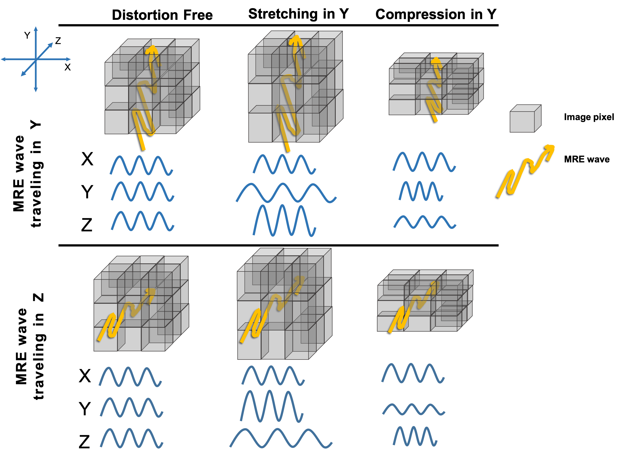
Geometric distortion in magnetic resonance imaging (MRI) is a common artifact arising from magnetic field inhomogeneity and susceptibility differences at air/tissue/bone interfaces[1]. While these distortions are well-characterized, and algorithms exist to correct these effects, the impact on quantitative MRI techniques, with or without correction, is generally underexamined. Magnetic resonance elastography (MRE) is a novel phase contrast MRI technique to calculate mechanical properties of soft tissue by imaging shear wave displacements and estimating stiffness through a nonlinear inversion algorithm. This inversion involves fitting measured wavefields to a numerical model of the elastodynamic wave problem, and thus geometric distortion in the measurements can lead to errors in estimated properties, particularly when wave propagation and distortion direction are different (Figure 1). In echoplanar imaging (EPI) sequences, distortion occurs in the phase encode direction and can be corrected to some degree via algorithms using a fieldmap, such as with FUGUE or TOPUP in FSL[2]. In high-resolution sequences, distortion is generally worse due to longer data readout times, however this can be counteracted through multishot sequences[3] with a tradeoff in longer scan time. As brain MRE development increasingly seeks to accurately map the properties of specific neuroanatomical regions[4], understanding the effect of distortion on quantitative measures is critical. In this study, we systematically examine quantitative effects of distortion and correction approaches on MRE data through a series of manipulated and realistic experiments.Methods
Distortion was characterized through simulations and in vivo brain experiments. A previously-reported, 3D finite-element simulation[5] based on separate gray and white matter stiffness was used. Simulation of k-space sampling used two type of trajectories: 2D EPI and spiral, manipulating distortion by changing readout time through bandwidth and number of interleaved spiral shots, respectively. All simulations resulted in the same image characteristics (2 mmisotropic resolution; FOV=100x100mm; 20 slices), and were iteratively reconstructed[6]. Linear ramp field inhomogeneity gradients were simulated and applied, with gradients between -80 to 80 Hz/cm, and a “realistic” fieldmap was also used that simulated field inhomogeneity near the frontal sinus cavity. EPI images were corrected using FUGUE in FSL, while spiral images were corrected using field correction during iterative reconstruction.
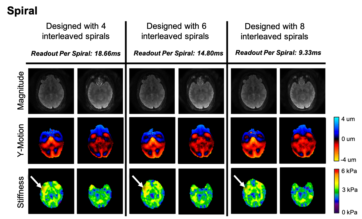
Three healthy adult subjects (1 male; ages 22-30) were imaged on a Siemens 3T Prisma scanner using a 64-channel head RF-receive coil. 50 Hz vibrations delivered to the head via a pneumatic actuator system with passive pillow driver (Resoundant, Inc.; Rochester, MN).MRE data was acquired using a 3D multiband, multishot spiral sequence7with 1.6 mm resolution and readout time was controlled via designed interleaved spiral shots (8, 6, and 4). Spiral data was reconstructed using [8]. All displacement images were processed using nonlinear inversion (NLI)[9] to estimate storage and loss modulus, G’ and G” respectively, then configured into viscoelastic shear stiffness, μ= 2|G|2/(G’+|G|).
Results and Discussion
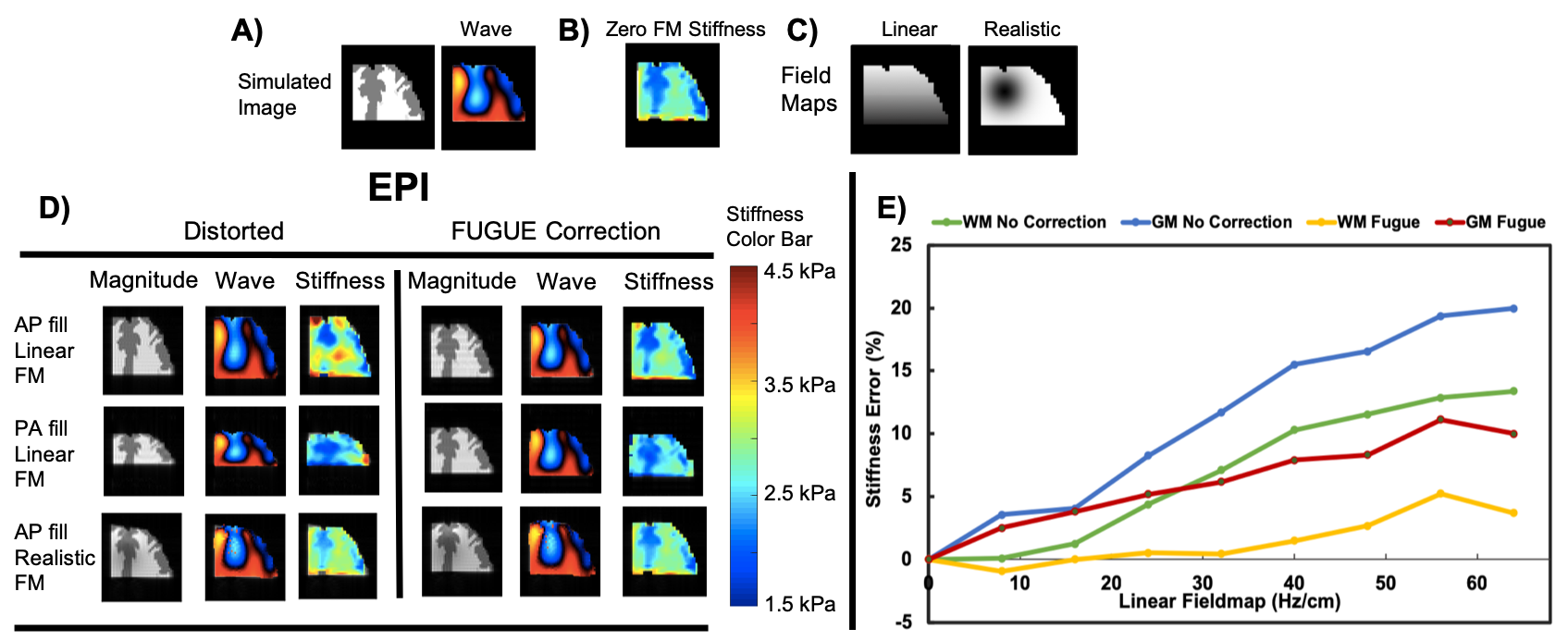
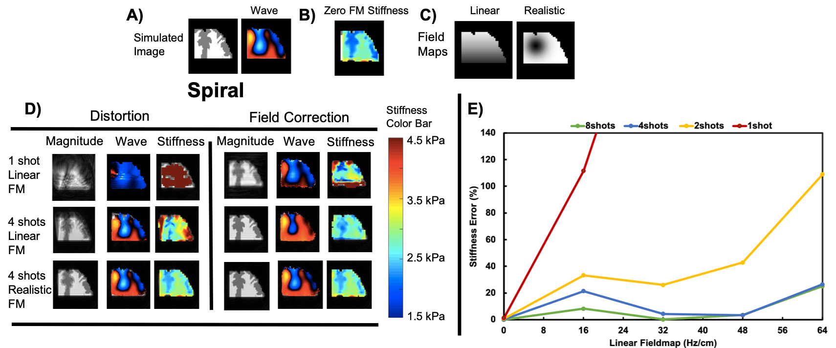
Directionally changing k-space filling of EPI sequences leads to an overestimate (anterior-posterior) or underestimate (posterior-anterior) of mechanical properties accompanied with a stretching or compressing of the image respectively (Figure 2). In both of these cases, using FSL FUGUE correction removes part, but not all of the error. For each data set, the level of distortion was calculated from the pixel shift in the phase encoding direction[1], ∆rpe(x,y,z) = γ∆B0(x,y,z)Tro, where γis gyromagnetic ratio, ∆B0 is the magnetic field inhomogeneity, and is the data readout time Tro. Distortion was determined as the gradient of the pixel shift map, d∆rpe/dy.Through our experiments, we have found that at typical MRE image readout times of 25 ms, 10% of the brain has waves images which are more than 5% distorted, and that image distortion leads to stiffness error in an exponential fashion such that 1% distortion leads to only 0.1% stiffness error, but 15% distortion leads to nearly 20% stiffness error. In spiral images, more accurate mechanical property calculations can be made with shorter readout times, controlled by increasing the number of interleaved spirals, with 4 interleaved spirals, or approximately a 22 ms readout time (Figure 3). Our in vivo spiral (Figure 5) experiments confirm our simulation findings and demonstrate a key consideration in designing high-resolution MRE acquisitions.Conclusion
To best develop MRE towards a clinical setting, sequences must be designed with high resolution and low geometric distortion. Any residual distortion will lead to mechanical property miscalculations. We have found through our study of distortion and distortion correction that accuracy limits do exist in MRE stiffness calculation in the presence of geometric distortion, and quantitative MRE studies should consider the effect of distortion when designing protocols.Acknowledgements
Delaware INBRE (P20-GM103446) and University of Delaware Research Foundation.References
[1] Jenkinson, M., Beckmann, C.F., Behrens, T.E.J., Woolrich, M.W., Smith, S.M., 2012. Fsl. Neuroimage 62, 782–790.
[2] Jezzard, P., Balaban, R.S., 1995. Correction for Geometric Distortion in Echo-Planar Images from B0 Field Variations. Magn. Reson. Med. 34, 65–73.
[3] CL Johnson, MDJ McGarry, EEW Van Houten, JB Weaver, KD Paulsen, BP Sutton, JG Georgiadis, “Magnetic Resonance Elastography of the Brain Using Multishot Spiral Readouts with Self-Navigated Motion Correction,” Magnetic Resonance in Medicine, 2013; 70(2):404-412.
[4] CL Johnson, EH Telzer, “Magnetic Resonance Elastography for Examining Developmental Changes in the Mechanical Properties of the Brain,” Developmental Cognitive Neuroscience, 2018; 33:176-181.
[5] MDJ McGarry, CL Johnson, BP Sutton, JG Georgiadis, EEW Van Houten, AJ Pattison, JB Weaver, KD Paulsen, “Suitability of Poroelastic and Viscoelastic Mechanical Models for High and Low Frequency MR Elastography,” Medical Physics, 2015; 42(2):947-957.
[6] Sutton, BP et al. IEEE Transactions on Medical Imaging, 2003
[7] CL Johnson, JL Holtrop, AT Anderson, BP Sutton, “Brain MR Elastography with Multiband Excitation and Nonlinear Motion-Induced Phase Error Correction,” 24th Annual Meeting of the International Society for Magnetic Resonance in Medicine, Singapore, May 7-13, 2016, p. 1951.
[8] Cerjanic, A. et al.PowerGrid: A open source library for accelerated iterative magnetic resonance image reconstruction. Proc Intl Soc Mag Reson Med14–17 (2016).
[9] Mcgarry, M.D.J., Houten, E.E.W. Van, Johnson, C.L., Georgiadis, J.G., Sutton, B.P., Weaver, J.B., Paulsen, K.D., 2012. Multiresolution MR elastography using nonlinear inversion.
Figures