3971
Fast Brain MR Elastography Using a Simultaneous Multislice EPI Acquisition on a Compact 3T Scanner1Radiology, Mayo Clinic, Rochester, MN, United States
Synopsis
In this study, we developed a fast MR elastography (MRE) pulse sequence using a simultaneous multislice (SMS)-EPI acquisition. The feasibility of SMS-EPI-MRE was demonstrated on a compact 3T scanner in a pilot volunteer study using 2 different head-coil arrays. Our results show that the overall scan time for 60-Hz 3-mm isotropic and 80-Hz 2-mm isotropic whole-brain MRE acquisitions were greatly reduced to 1-2 minutes (depending on the multiband acceleration) and 3:21 minutes, respectively. The wave images and stiffness maps from the SMS acquisitions were comparable to conventional MRE results.
Introduction
Brain MR elastography (MRE) is an emerging noninvasive imaging technique that has been widely used to detect diseased or degenerated tissue by imaging differences in shear wave propagation associated with altered tissue stiffness1. When applied to the brain it is typically desired to perform MRE with higher frequencies and higher spatial resolution. However, the trade-off is often longer scan times. With fast imaging sequences such as EPI or spiral, an 8-phase-offset whole-brain MRE exam may still require ~3-7 minutes for a 3-mm isotropic resolution scan2,3, and ~6-10 minutes for a 2-mm isotropic resolution scan4,5. Recently, the simultaneous multislice (SMS) technique has been widely used in fMRI and diffusion MRI to significantly reduce the scan time with little impact on SNR6. Although earlier studies demonstrated the combination of MRE with SMS7,8, those acquisitions were based on gradient-recalled echo (GRE) or spiral sequence rather than the more commonly used EPI-based sequence for clinical brain MRE. Therefore, the purpose of this study was to develop an SMS-EPI-MRE pulse sequence and demonstrate its feasibility to significantly accelerate the acquisition of brain MRE data on a compact 3T scanner while maintaining good image quality, SNR, and stiffness fidelity.Method
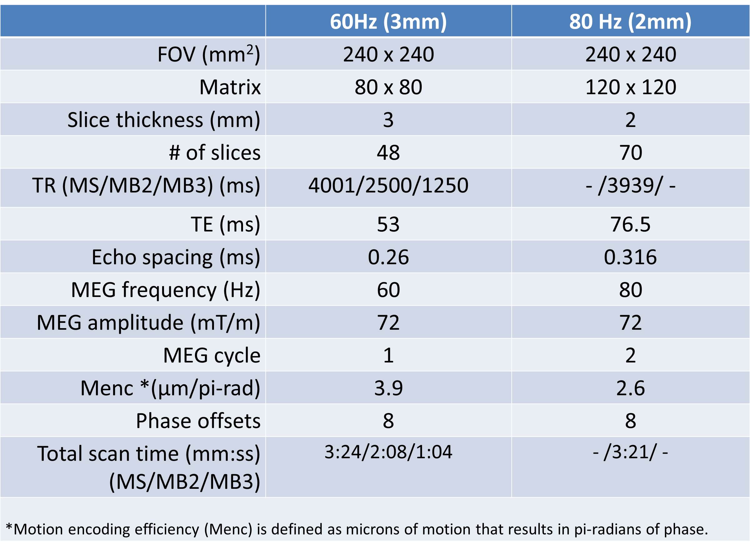
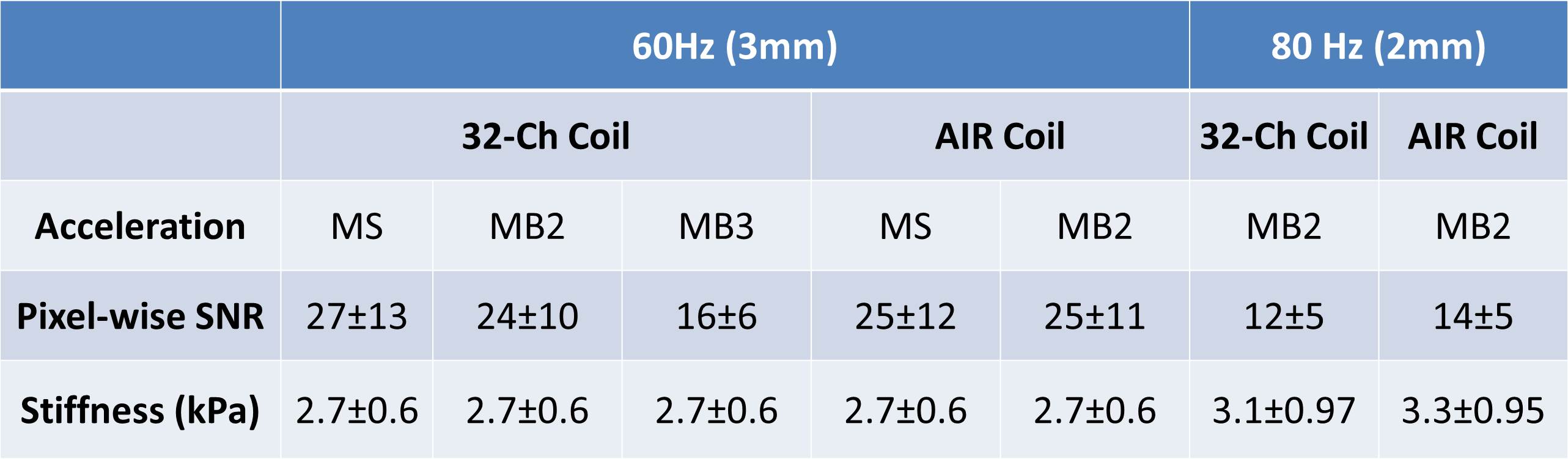
Multiband (MB) RF pulses and blipped-CAIPI9 slice-dependent phase modulation were implemented into a single-shot, spin-echo (SE), EPI-MRE sequence on a compact 3T head-only scanner with a high-performance gradient coil (maximum gradient amplitude of 80 mT/m and slew rate of 700 T/m/s)10-14. One volunteer was scanned using a 32-channel (32-Ch) head coil (Nova Medical, Wilmington, MA) and a 16-channel flexible adaptive image receive (AIR) head coil built in-house with GE (GE Healthcare, Waukesha, WI ) AIR® coil elements15. The 60-Hz MRE data were acquired with a 3-mm isotropic resolution and whole-brain coverage using the conventional multislice (MS) scheme and SMS with a MB acceleration factor of 2 (MB2) and 3 (MB3). The TR was reduced for MB2 and MB3 to the minimum necessary to perform all of the needed slice excitations. The total scan time for 6 motion-encoding directions and 8 phase offsets was 3:24, 2:08 and 1:04 minutes for MS, MB2, and MB3, respectively. The 80-Hz whole-brain MRE data were acquired with a 2-mm isotropic resolution using MB2. To increase the motion sensitivity, two cycles of the motion-encoding gradient (MEG) were applied on each side of the 180° RF pulse, leading to a total scan time of 3:21 minutes. The details of the scan parameters are provided in Table 1. The pixelwise magnitude SNR was calculated by taking the ratio of the mean and standard deviation of each pixel over the 48 repeatedly acquired volumes for each experiment. The stiffness maps were generated using a recently developed artificial neural network inversion (NNI) algorithm16.Results
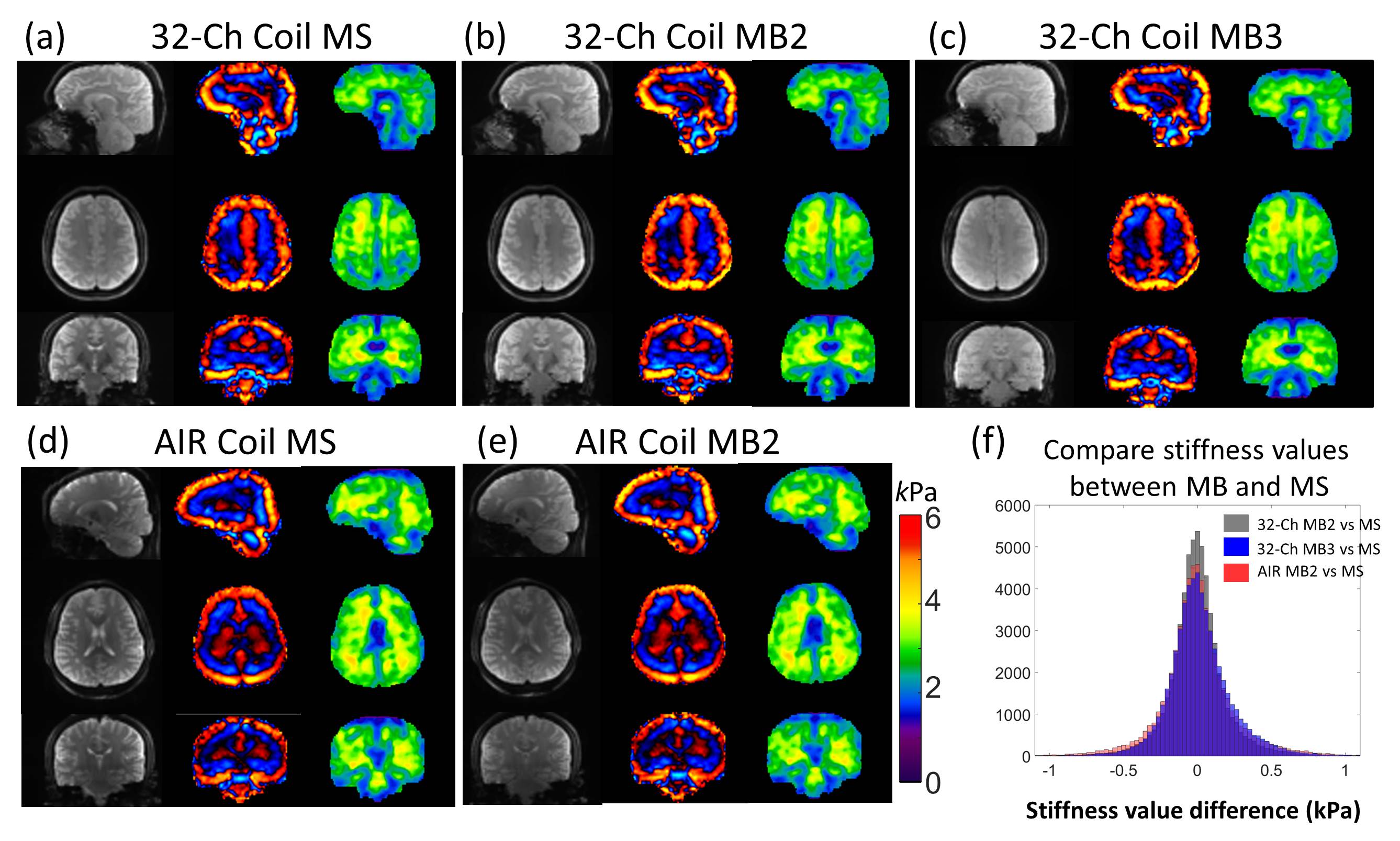
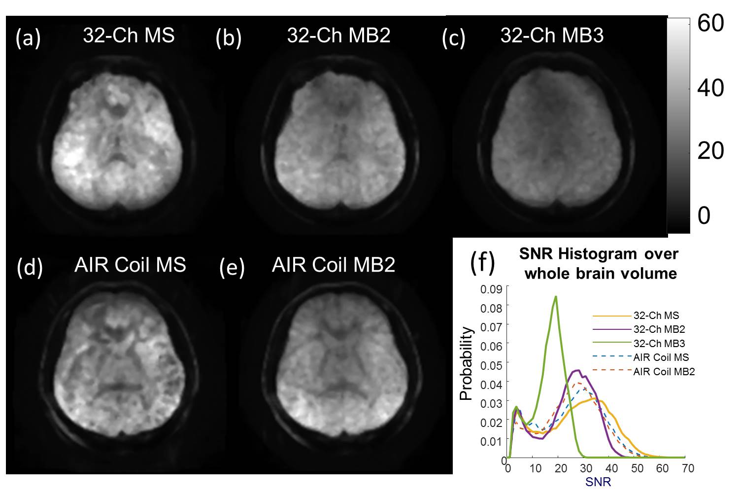
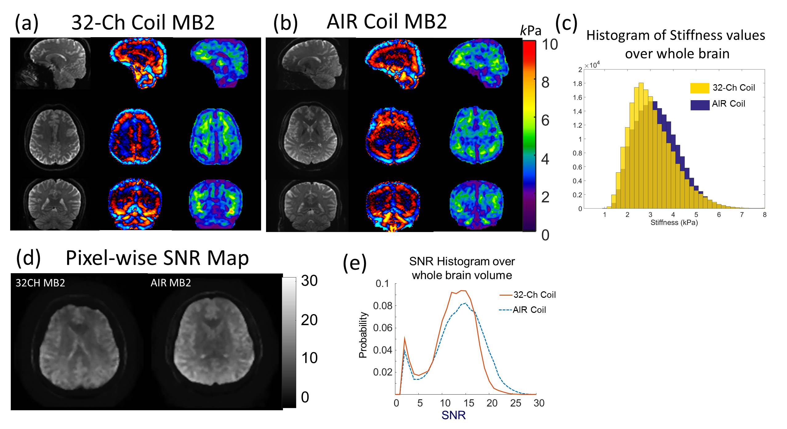
A comparison of the results from the SMS and conventional MRE acquisitions is shown in Figure 1. The MB2 and MB3 acquisitions greatly reduced the overall scan time by 37% and 69%, respectively, with the acquired wave images visually identical to those from the conventional acquisition. No significant difference was found in the whole-brain average stiffness from the different acquisitions (Table 2). Figure 2 shows a representative slice of the pixelwise SNR maps for the 60-Hz acquisitions as well as the SNR histograms for the whole-brain volume. As expected, the MB3 SNR was significantly lower than the other scans (Table 2) due to the signal saturation with a short TR (1250 ms). The 80-Hz MRE results acquired with MB2 on the 32-channel head coil and the AIR head coil are shown in Figure 3. The AIR coil exhibited higher magnitude SNR but slightly lower wave amplitude, which may be due to the different coupling between the MRE passive driver and the AIR coil.Discussion and Conclusion
We have demonstrated the SMS technique is compatible with SE-EPI brain MRE using a compact 3T scanner and both a 32-channel head coil and a 16-channel AIR coil array. The SMS implementation significantly reduces the overall scan time (e.g., 1-2 minutes for the 60-Hz MRE acquisition depending on the multiband acceleration). Combined with the soft and flexible AIR coil, SMS-EPI-MRE may greatly improve patient comfort. Although the magnitude SNR for MB3 was lower due to the short TR used, the wave SNR was sufficient to reliably estimate stiffness using the NNI method, which has been proven to be more robust to noise compared to conventional direct-inversion methods16. Furthermore, given the high-performance gradient performance of the compact 3T scanner, a 2-mm, isotropic, 80-Hz, whole-brain MRE scan can be achieved in under 3.5 minutes with sufficient SNR for the neural-network MRE inversion, facilitating MRE studies focusing on smaller regions across the brain.Acknowledgements
This work was supported in part by research grant NIH U01-EB024450References
1 Murphy, M. C., Huston, J., 3rd & Ehman, R. L. MR elastography of the brain and its application in neurological diseases. Neuroimage, (2017).
2 Hughes, J. D., Fattahi, N., Van Gompel, J., Arani, A., Meyer, F., Lanzino, G., Link, M. J., Ehman, R. & Huston, J. Higher-Resolution Magnetic Resonance Elastography in Meningiomas to Determine Intratumoral Consistency. Neurosurgery 77, 653-658, (2015).
3 Yin, Z., Sui, Y., Trzasko, J. D., Rossman, P. J., Manduca, A., Ehman, R. L. & Huston, J., 3rd. In vivo characterization of 3D skull and brain motion during dynamic head vibration using magnetic resonance elastography. Magn Reson Med, (2018).
4 Johnson, C. L., McGarry, M. D., Van Houten, E. E., Weaver, J. B., Paulsen, K. D., Sutton, B. P. & Georgiadis, J. G. Magnetic resonance elastography of the brain using multishot spiral readouts with self-navigated motion correction. Magn Reson Med 70, 404-412, (2013).
5 Johnson, C. L., Holtrop, J. L., McGarry, M. D. J., Weaver, J. B., Paulsen, K. D., Georgiadis, J. G. & Sutton, B. P. 3D Multislab, Multishot Acquisition for Fast, Whole-Brain MR Elastography with High Signal-to-Noise Efficiency. Magnetic Resonance in Medicine 71, 477-485, (2014).
6 Barth, M., Breuer, F., Koopmans, P. J., Norris, D. G. & Poser, B. A. Simultaneous multislice (SMS) imaging techniques. Magn Reson Med 75, 63-81, (2016).
7 Guenthner, C., Runge, J. H., Sinkus, R. & Kozerke, S. Simultaneous Multislice Acquisition for Magnetic Resonance Elastography. ISMRM 25, 1132, (2017).
8 Johnson, C. L., Holtrop, J. L., Anderson, A. T. & Sutton, B. P. Brain MR elastography with multiband excitation and nonlinear motion-induced phase error correction. ISMRM 24, 1951, (2016).
9 Setsompop, K., Gagoski, B. A., Polimeni, J. R., Witzel, T., Wedeen, V. J. & Wald, L. L. Blipped-controlled aliasing in parallel imaging for simultaneous multislice echo planar imaging with reduced g-factor penalty. Magn Reson Med 67, 1210-1224, (2012).
10 Weavers, P. T., Shu, Y. H., Tao, S. Z., Huston, J., Lee, S. K., Graziani, D., Mathieu, J. B., Trzasko, J. D., Foo, T. K. F. & Bernstein, M. A. Technical Note: Compact three-tesla magnetic resonance imager with high-performance gradients passes ACR image quality and acoustic noise tests. Medical Physics 43, 1259-1264, (2016).
11 Foo, T. K. F., Laskaris, E., Vermilyea, M., Xu, M., Thompson, P., Conte, G., Van Epps, C., Immer, C., Lee, S. K., Tan, E. T., Graziani, D., Mathieu, J. B., Hardy, C. J., Schenck, J. F., Fiveland, E., Stautner, W., Ricci, J., Piel, J., Park, K., Hua, Y., Bai, Y., Kagan, A., Stanley, D., Weavers, P. T., Gray, E., Shu, Y., Frick, M. A., Campeau, N. G., Trzasko, J., Huston, J., 3rd & Bernstein, M. A. Lightweight, compact, and high-performance 3T MR system for imaging the brain and extremities. Magn Reson Med 80, 2232-2245, (2018).
12 Weavers, P. T., Tao, S., Trzasko, J. D., Frigo, L. M., Shu, Y., Frick, M. A., Lee, S. K., Foo, T. K. & Bernstein, M. A. B0 concomitant field compensation for MRI systems employing asymmetric transverse gradient coils. Magn Reson Med, (2017).
13 Tao, S. Z., Weavers, P. T., Trzasko, J. D., Shu, Y. H., Huston, J., Lee, S. K., Frigo, L. M. & Bernstein, M. A. Gradient Pre-Emphasis to Counteract First-Order Concomitant Fields on Asymmetric MRI Gradient Systems. Magnetic Resonance in Medicine 77, 2250-2262, (2017).
14 Tao, S., Trzasko, J. D., Gunter, J. L., Weavers, P. T., Shu, Y., Huston, J., Lee, S. K., Tan, E. T. & Bernstein, M. A. Gradient nonlinearity calibration and correction for a compact, asymmetric magnetic resonance imaging gradient system. Phys Med Biol 62, N18-N31, (2017).
15 McGee, K. P., Stormont, R. S., Lindsay, S. A., Taracila, V., Savitskij, D., Robb, F., Witte, R. J., Kaufmann, T. J., Huston, J., Riederer, S. J., Borisch, E. A. & Rossman, P. J. Characterization and evaluation of a flexible MRI receive coil array for radiation therapy MR treatment planning using highly decoupled RF circuits. Physics in Medicine and Biology 63, (2018).
16 Murphy, M. C., Manduca, A., Trzasko, J. D., Glaser, K. J., Huston, J., 3rd & Ehman, R. L. Artificial neural networks for stiffness estimation in magnetic resonance elastography. Magn Reson Med 80, 351-360, (2018).
Figures