3966
Online platform for extendable server-based processing of magnetic resonance elastography data.1Department of Radiology, Charité - Universitätsmedizin Berlin, Berlin, Germany, 2Insitute of Medical Informatics, Charité - Universitätsmedizin Berlin, Berlin, Germany, 3Department of Radiology, The Ohio State University, Columbus, OH, United States
Synopsis
The variety of reconstruction algorithms throughout the MRE community makes it difficult to compare the validity and strengths of these methods. We propose an extendable online platform, combining multiple processing methods to facilitate a direct comparison. The platform is implemented as a Java web application, designed to run every command line program. Processing is done on the server, configured through the easy-to-use web-interface. Currently, state-of-the-art MRE reconstruction algorithms, multiple pre-processing methods, automated data import as well as sample data of phantom-MRE and in-vivo MRE are included. We aim at extension of the platform and an automation of adding new methods.
Introduction
Magnetic
resonance elastography (MRE) is an important clinical method for staging
hepatic fibrosis1. Further applications of MRE for the
diagnosis of degenerative, inflammatory or oncological diseases are under
development or are currently being validated in clinical trials2. Besides hardware and sequences, an
important technical component of MRE are reconstruction algorithms, which
convert wave images into maps of elasticity- and viscosity-related parameters3. Worldwide, research groups
continuously improve existing methods or develop new approaches based on new
mathematical concepts in the field of inverse-problem solutions4. However, the variety of
reconstruction approaches throughout the MRE community makes it difficult to
compare the validity and strengths of new methods between groups and
applications. Therefore, we propose an open online platform facilitating a
direct comparison by combining multiple methods on one platform. By
implementing the platform as a web application, updates of the methods are
automatically maintained and worldwide distributed, assuring that consistently the
newest version is used.Methods
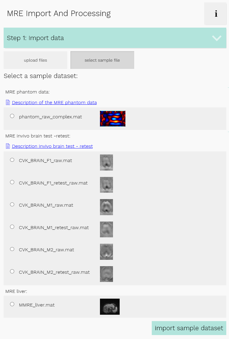
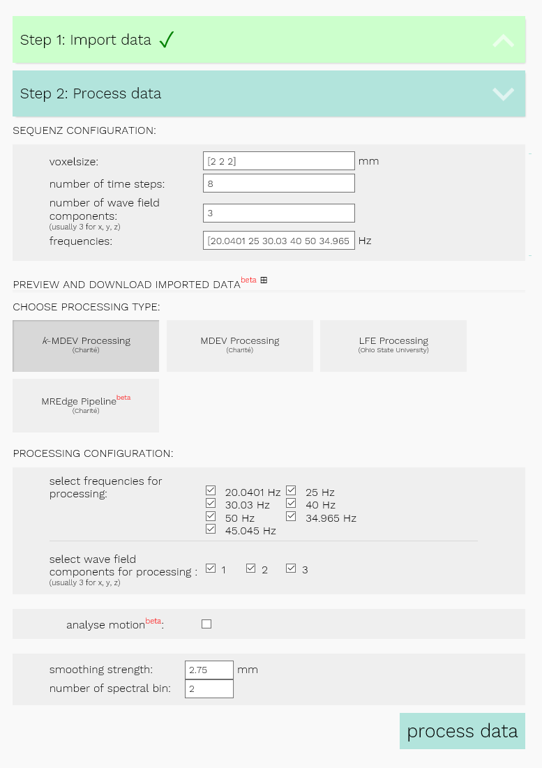
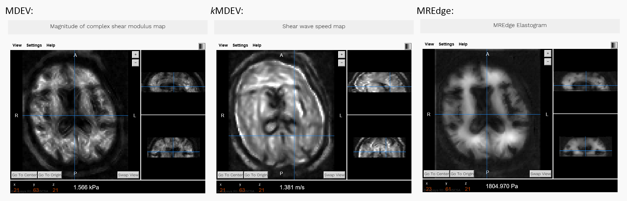
The online platform is implemented as a Java web application running on a scalable virtual server (https://bioqic-apps.charite.de). The web-interface is designed for uploading and importing data (figure 1), configuring the processing pipelines (figure 2) and receiving the results (figure 3). All processing is done on the server side. The platform is designed to be able to run every command line program that can be executed on an Ubuntu 16.04 system. Currently compiled MATLAB programs are executed for the import and the processing pipelines. Using asynchronous communication between client and server, the content of the web-interface can be dynamically updated depending on the processing step. This ensures a clear and easy-to-use user-interface, allowing also for displaying real-time progress information parsed from the output of the algorithms during execution. Communication between the client and the server is secured by SSL encryption using the HTTPs protocol. All data uploaded by users will be automatically deleted after logging out or after 1 h of inactivity. By using the Papaya medical research image viewer5, the results can be directly displayed as a preview on the web-interface (figure 3).Results
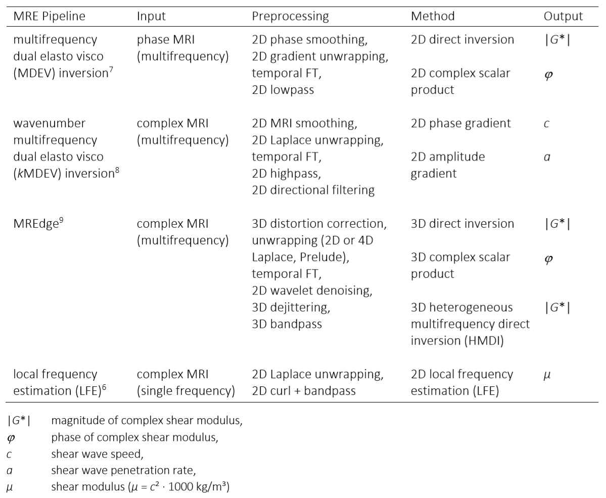
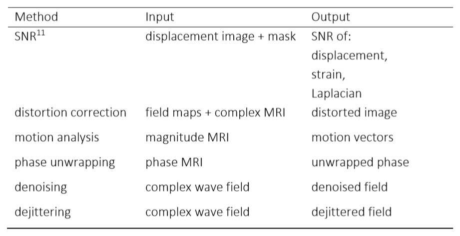
Currently the platform offers a widely automated import of data in DICOM, NIfTI, and MATLAB format. State-of-the-art image reconstruction pipelines for MRE data (figure 4), as well as preprocessing methods for phase unwrapping, wavelet denoising, dejittering and motion correction are provided (figure 5). The latter can be used either in combination with the inversion pipelines or as standalone applications. The configuration of these pipelines can be easily done through the web-interface with predefined presets or manually. Program execution starts with an upload of data or the selection of a sample data (figure 1). These are then automatically imported. Next, if not automatically detected, MRE related parameters have to be specified. Subsequently a processing pipeline can be chosen (figure 2). So far, pipelines are available for a variety of inversion methods including LFE6, MDEV7, kMDEV8, and HMDI9 (figure 4). Additionally, multiple preprocessing steps such as different unwrapping methods, dual-tree complex wavelet denoising10 or bandpass filtering can be combined with 2D or 3D inversion methods or run as their own applications on uploaded data (figure 5). Results as well as the imported data, can be viewed on the web-interface or are available as download in MATLAB, DICOM and NIfTI format.Discussion
To date, the implemented platform is the only web-based application for processing of MRE data. The provided state-of-the-art postprocessing and inversion methods can easily be accessed and compared in an unified platform by all research groups interested in MRE. Since processed data can be downloaded in multiple formats, the platform facilitates easy comparison with own methods locally. The software architecture allows a straightforward implementation of new methods and extension of all processing options including data repository.Conclusion
The proposed extendable server-based processing of MRE data facilitates comparison of data processing pipelines in MRE on own data and standardized sample data. The platform is continuously maintained and can be extended by including new data processing methods. Feedback from users of different research fields is included in improvements and upgrades to make the platform more user-friendly and to improve its functionality. We are aiming to include more data processing pipelines, also from other quantitative imaging methods such as arterial spin labelling, 4D-flow or diffusion MRI to support multiparametric data analysis. This will be accompanied by an expansion of the data repository. Finally, we want to offer an automated process for the integration of new methods so that other research groups worldwide can participate in the implementation in own pipelines more easily.Acknowledgements
This research was supported by GRK2260 (BIOQIC) of the German Research Foundation.References
1. Venkatesh SK, Yin M, Ehman RL. Magnetic resonance elastography of liver: Technique, analysis, and clinical applications. J Magn Reson Imaging 2013; 37(3): 544-55.
2. Dong H, White RD, Kolipaka A. Advances and Future Direction of Magnetic Resonance Elastography. Top Magn Reson Imaging 2018; 27(5): 363-84.
3. Hirsch S, Braun J, Sack I. Magnetic Resonance Elastography: Physical Background And Medical Applications: Wiley-VCH; 2017.
4. Doyley MM. Model-based elastography: a survey of approaches to the inverse elasticity problem. Phys Med Biol 2012; 57(3): R35-73.
5. University of Texas Health Science Center. Papaya. 2018. http://rii-mango.github.io/Papaya/ (accessed 06.11.2018).
6. Manduca A, Oliphant TE, Dresner MA, et al. Magnetic resonance elastography: non-invasive mapping of tissue elasticity. Med Image Anal 2001; 5(4): 237-54.
7. Hirsch S, Guo J, Reiter R, et al. MR Elastography of the Liver and the Spleen Using a Piezoelectric Driver, Single-Shot Wave-Field Acquisition, and Multifrequency Dual Parameter Reconstruction. Magn Reson Med 2014; 71(1): 267-77.
8. Tzschatzsch H, Guo J, Dittmann F, et al. Tomoelastography by multifrequency wave number recovery from time-harmonic propagating shear waves. Med Image Anal 2016; 30: 1-10.
9. Barnhill E, Davies PJ, Ariyurek C, Fehlner A, Braun J, Sack I. Heterogeneous Multifrequency Direct Inversion (HMDI) for magnetic resonance elastography with application to a clinical brain exam. Med Image Anal 2018; 46: 180-8.
10. Barnhill E, Hollis L, Sack I, et al. Nonlinear multiscale regularisation in MR elastography: Towards fine feature mapping. Med Image Anal 2016; 35: 133-45.
11. Bertalan G, Guo J, Tzschatzsch H, et al. Fast tomoelastography of the mouse brain by multifrequency single-shot MR elastography. Magn Reson Med 2018.
Figures