3963
Tabletop MR Elastography (MRE): Preliminary Results Towards an Assessment of Frozen Tissue Bank Samples.1Bioengineering, University of Illinois at Chicago, Chicago, IL, United States, 2Radiology, Charité – Universitätsmedizin Berlin, corporate member of Freie Universität Berlin, Humboldt-Universität zu Berlin, and Berlin Institute of Health, Berlin, Germany
Synopsis
Tabletop magnetic resonance elastography (MRE) at 0.5 Tesla was recently introduced as a low-cost technique for assessing mechanical properties of small tissue samples. Frozen tissue banks potentially facilitate access to large amounts of specimens. Therefore, we aimed to characterize changes
Introduction
Tabletop magnetic resonance elastography (MRE) at 0.5 Tesla was recently introduced as a low-cost technique for assessing mechanical properties of small tissue specimens.1–3 Meanwhile, frozen tissue banks are progressively emerging, and moving into clinical care due to high quality preservation of biological tissues compared to conventional formalin-based fixation.4 Frozen tissue banks potentially facilitate access to large amounts of well-preserved specimens for tabletop MRE. Therefore, we aimed to characterize changes of mechanical properties of fresh ex vivo (native), and frozen and thawed (lysed) porcine muscle, liver and kidney specimens.Methods
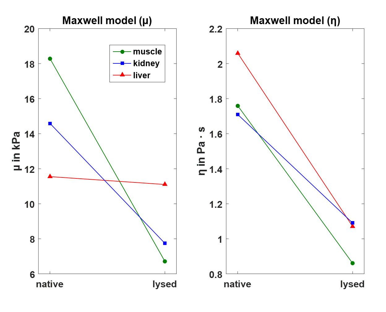
MRE was performed using a 0.5 Tesla tabletop magnetic resonance imaging scanner (DC 600, Pure Devices GmbH, Würzburg, Germany) and a custom-build piezo-electric actuator at four drive frequencies (500, 1000, 1500, 2000 Hz). MRE data was acquired with a matrix of 128 x 128, field of view of 9.6 x 9.6 mm and 5 mm slice thickness. A geometrical focusing approach was employed to compensate for the changes in wavelength and attenuation over the range of frequencies. The shear modulus was calculated at each frequency using a fitting procedure of the displacement function for the cylindrical experimental setup used, which is based on Bessel functions.5 Specimens from porcine lumbar muscle (n = 3), porcine liver (n = 3) and porcine kidney (n = 3) were placed in a glass capillary and scanned twice: i) immediately after obtained from a butcher shop (native) ii) Afterward, specimens were stored at -20°C for 24 hours and rescanned after complete thawing (lysed). The rheological Maxwell model was fitted to obtain frequency independent tissue properties. Maxwell rheological parameters are µ (kPa) for stiffness and η (Pa · s) for viscosity.Results
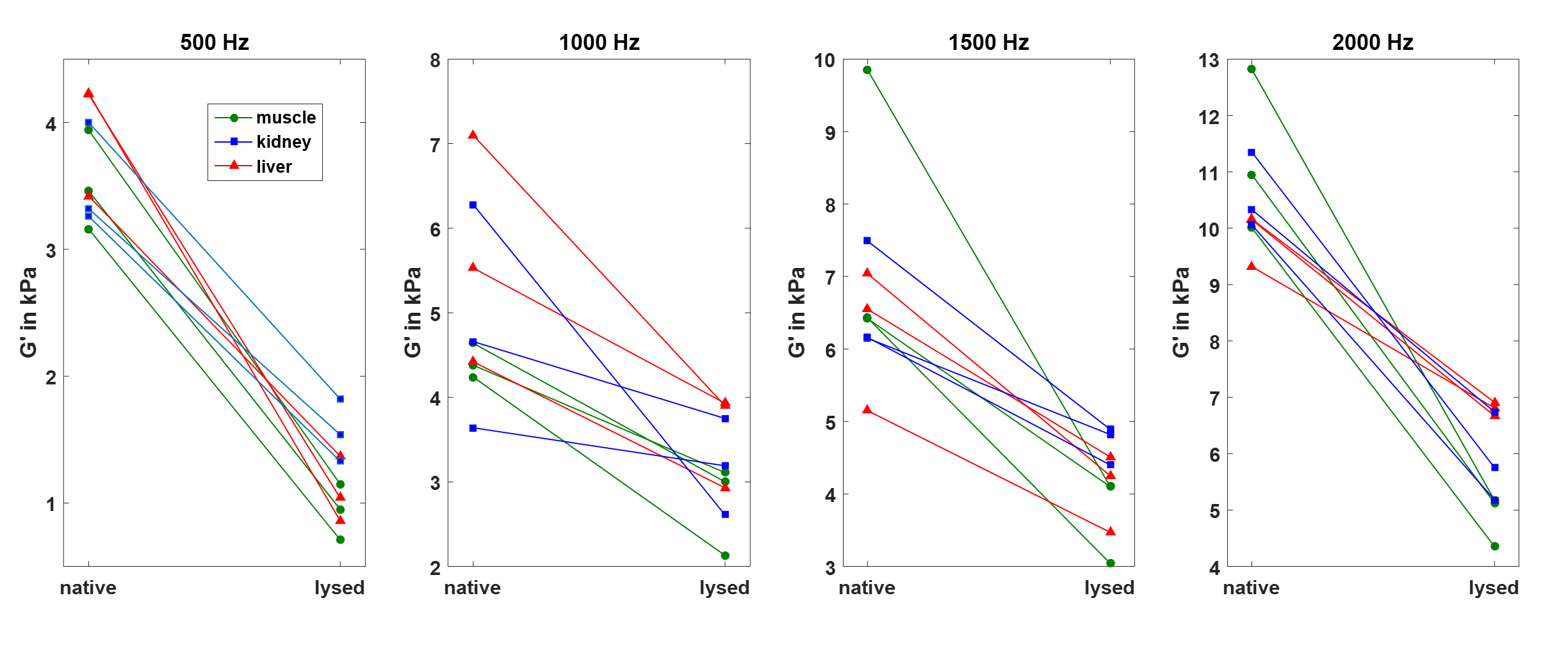
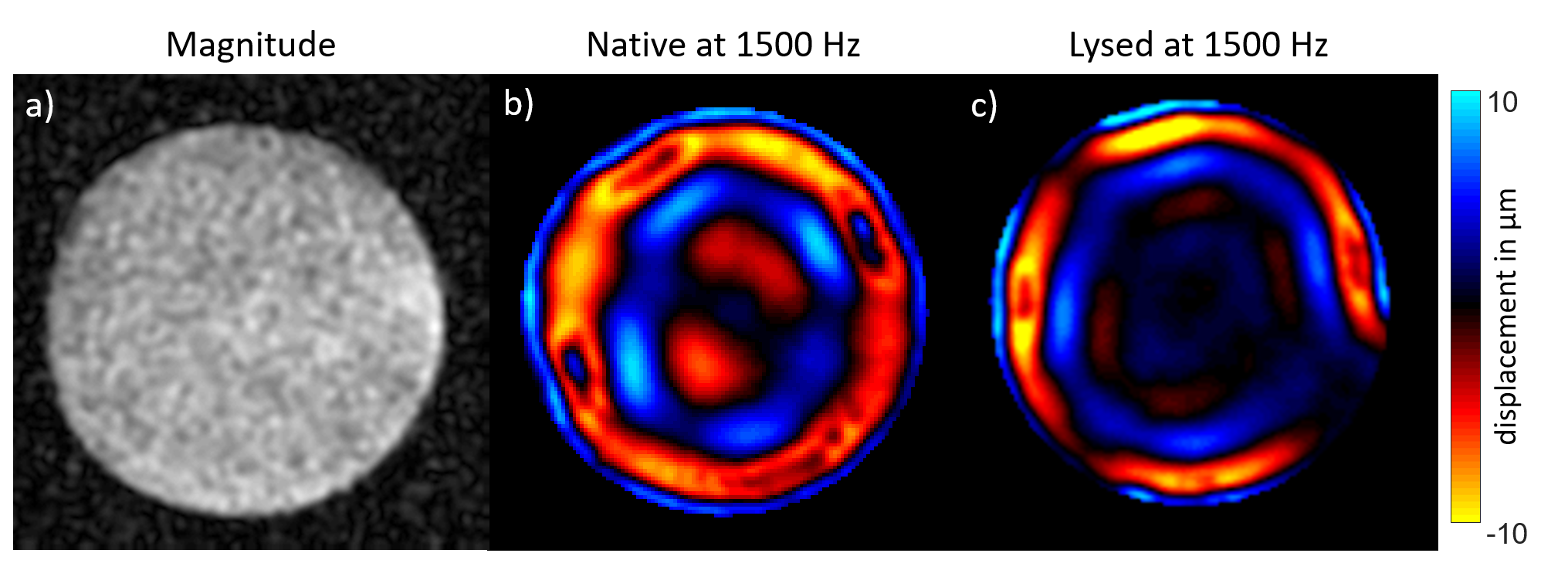
After freezing and complete thawing, a decrease in stiffness (real part G’ of the complex modulus) for all tissues and frequencies was evident as shown in figure 1. It was already perceptible by visual inspection of the shear wave images that shear wave length decreased in lysed tissue, which indicated reduced stiffness. An example image is illustrated in figure 2 for a kidney tissue sample at 1500 Hz. The ratio of mean G’ for lysed/native muscle, liver and kidney was 0.46, 0.57 and 0.60, respectively. A two-way analysis of variance (ANOVA) showed a statistical difference between mean values of all native and all lysed tissues with a P-value < 0.0001. Frequency independent mean values for µ and η (± standard deviation) derived from the Maxwell model for native and lysed tissues were as follows: muscle, 18.29 (2.57) and 6.71 (0.04) kPa, 1.76 (0.22) and 0.86 (0.12) Pa · s; liver, 11.55 (3.02) and 11.10 (2.10) kPa, 2.06 (0.43) and 1.07 (0.04) Pa · s; kidney, 14.59 (1.14) and 7.74 (1.27) kPa, 1.71 (0.21) and 1.09 (0.07) Pa · s. Additionally, mean values for µ and η for different tissues were displayed in figure 3. The decrease of mean values of both µ and η for muscle (ratio lysed/native: µ, 0.37 and η, 0.49) and kidney (ratio lysed/native: µ, 0.53 and η, 0.64) indicated a tissue softening after lysis. For liver, only a decrease in η was observed while µ decreased less pronounced (ratio lysed/native: µ, 0.96 and η, 0.52).Discussion
The uniform decrease of stiffness for single frequencies of lysed tissues is in alignment with a recent study investigating rat liver tissue.3 It is assumed that this effect is based on a damaged cell wall integrity of frozen tissue. However, after rheological fitting of the Maxwell model, our results showed a less pronounced stiffness decrease for liver tissue compared to muscle and kidney tissue. Evidence about changes of rheological tissue properties through lysis is currently limited. Therefore, applicability and accuracy of rheological models for fitting data from lysed tissues remain uncertain. The investigation of more specimens is planned.Conclusion
This study provides motivation for further investigation of frozen tissue bank samples using tabletop MRE as a low-cost technique for the assessment of mechanical tissue properties of small specimens. Moreover, this study motivates the evaluation and comparison of different rheological models for native and lysed biological tissues.Acknowledgements
Support by the 2016 Seed and Annual Funds of the College of Engineering at the University of Illinois at Chicago is kindly acknowledged.
Support from the Deutsche Forschungsgemeinschaft (DFG, German Research Foundation) is kindly acknowledged – RE 4161/1-1 (Rolf Otto Reiter).
References
1. Ipek-Ugay S, Drießle T, Ledwig M, et al. Tabletop magnetic resonance elastography for the measurement of viscoelastic parameters of small tissue samples. J Magn Reson. 2015;251:13–18.
2. Braun J, Tzschätzsch H, Körting C, et al. A compact 0.5T MR elastography device and its application for studying viscoelasticity changes in biological tissues during progressive formalin fixation. Magn Reson Med. 2017;00:1–9.
3. Angela Ariza de Schellenberger, Hannah Everwien, Nils Haep, et al. Mechanical characterization of rat liver tissue in native, lysed and decellularized states by 0.5 T tabletop magnetic resonance elastography (MRE). Proc Intl Soc Mag Reson Med. 2018;(5588).
4. Shabihkhani M, Lucey GM, Wei B, et al. The procurement, storage, and quality assurance of frozen blood and tissue biospecimens in pathology, biorepository, and biobank settings. Clin Biochem. 2014;47(4–5):258–66.
5. Yasar TK, Royston T, Magin RL. Wideband MR elastography for viscoelasticity model identification. Magn Reson Med. 2013;70:479–489.
Figures