3961
The contribution of cells and extracellular matrix to liver viscoelasticity: Histological analysis combined with multifrequency MR elastography and diffusion weighted imaging in rat liver specimens1Radiology, Charité-Universitätsmedizin Berlin, Berlin, Germany, 2University of Pennsylvania, Philadelphia, PA, United States
Synopsis
The effect of freezing/thawing at -20°C and -80°C on liver tissue was analyzed by diffusion-weighted imaging (DWI), multifrequency MR elastography (MRE) and histological methods. Freezing-induced deterioration of cell membranes, detachment of sinusoidal endothelial cells and disorganization of sinusoidal collagen together with reduced water diffusion and macroscopic shear modulus and affected tissue viscosity. Histological analysis revealed that collagen structures at the space of Disse are better preserved at -80°C than -20°C which was mirrored by tissue viscosity indicating the sensitivity of multifrequency MRE to microstructural changes of soft biological tissues.
Introduction
Little is known about the relationship between the liver's microarchitecture and macroscopic biophysical imaging markers such as water diffusion or shear viscoelasticity[1] as measured by diffusion-weighted imaging (DWI)[2] or MR elastography (MRE)[3]. Therefore, we modulated the integrity of liver parenchyma by freezing/thawing procedures and applied DWI and MRE combined with histological analyses. Although freezing of soft biological tissues leads to significant palpable tissue deteriorations, the underlying micro-structural changes have not been systematically studied[4]. Understanding the effect of freezing on the microarchitecture of liver tissue and identifying preservation-related tissue changes based on histological analyses and MRE examinations is relevant for biomedical research and regenerative medicine[5].Methods
Six groups of 44 liver specimen from Wistar rats (3 to 4 months) were investigated: native tissue (MRE:n=11, DWI:n=6), -20°C-frozen/thawed tissue (MRE:n=11, DWI:n=6), -80°C-frozen/thawed tissue (MRE:n=6, DWI:n=6). Cylindrical samples of 10mm height and 8mm diameter were chopped from the freshly harvested livers and frozen overnight at -20° or -80°C.
Histology
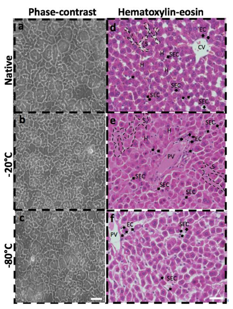
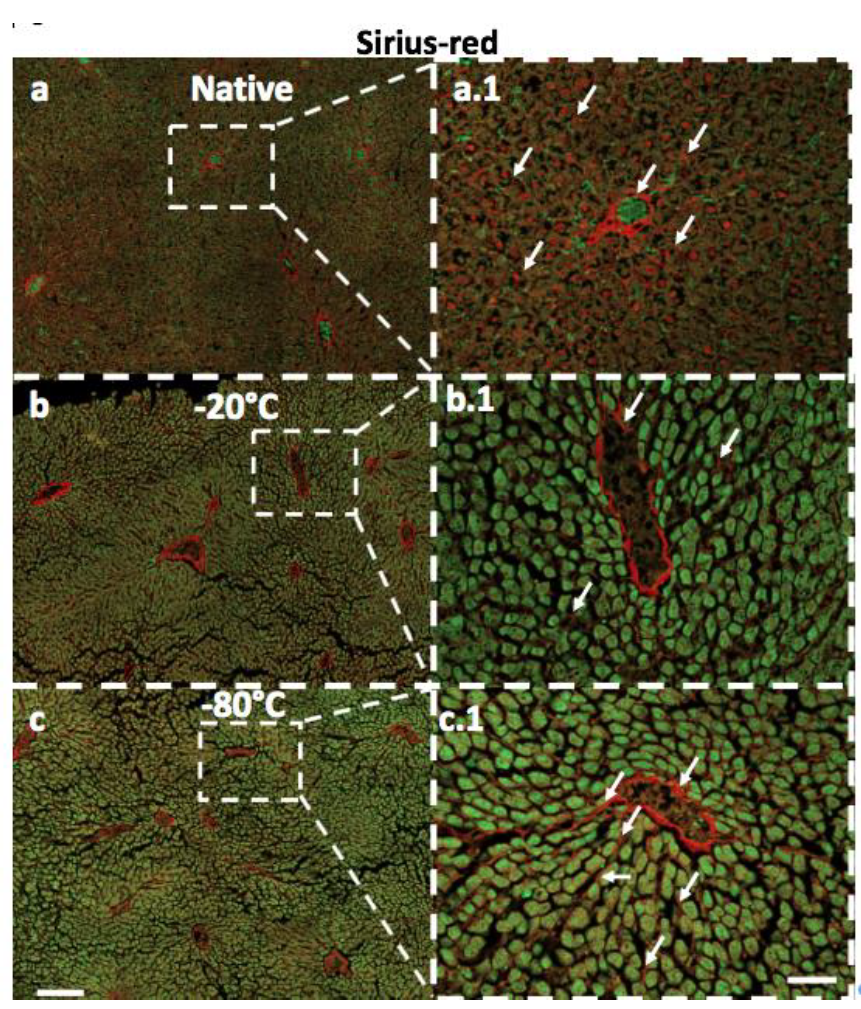
Liver cryosections were prepared for phase-contrast microscopy from unfixed liver tissue snap-frozen in precooled (-20°C) 2-methylbutane on top of dry ice and imbedded in cryomolds and stored at -20°C until sections were cut (5µm) in a microtom. Liver tissue fixed for 24h in 4% paraformaldehyde was dehydrated in ethanol and xylol. Standardized protocols were used for Harris Hematoxylin-eosin (HE), Masson’s Trichrome (M.T.) and Sirius red (S.R.) staining.
MRE and DWI
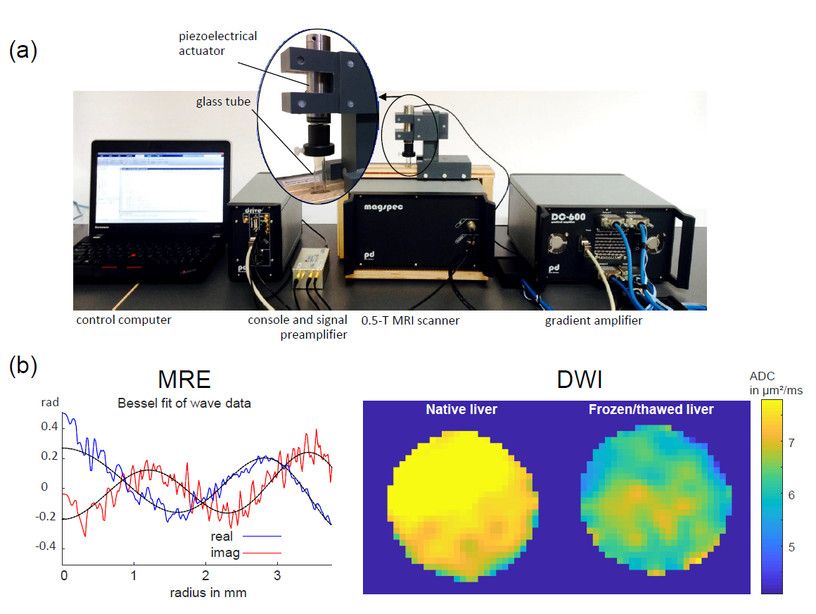
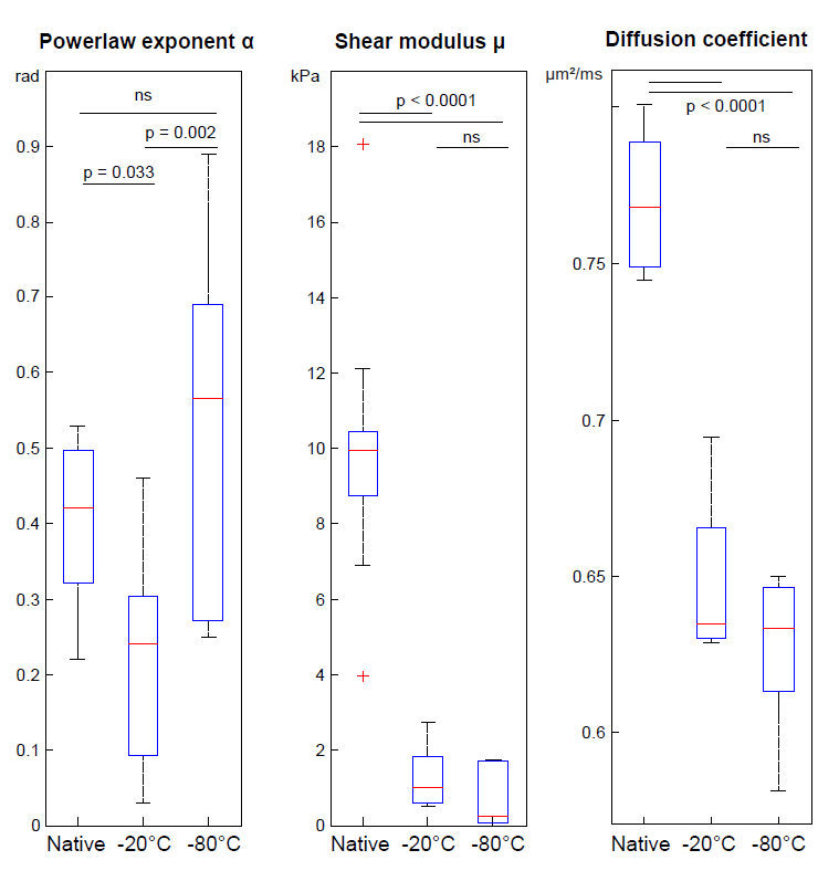
0.5-T tabletop MRI (PureDevices, Germany) was used for MRE and DWI. For MRE, a customized spin-echo sequence and a piezoelectric driver were used as detailed in[6] and illustrated in Fig.1a. The range of vibration frequencies was 300-800 Hz with 100-Hz-increments. Further imaging parameters: TR=2000ms, TE=42ms, 8 wave dynamics, single slice of 3mm thickness, matrix size=64x64, field-of-view=9.6x9.6mm². Post processing was based on Bessel fits as described previously[6] (Fig.1b) yielding one shear-wave speed c per frequency. The frequency dispersion of c was then fitted by a two-parameter powerlaw according to the springpot-model yielding shear modulus μ and powerlaw exponent α[7]. For DWI, a customized spin-echo sequence with one pair of split diffusion gradients was applied to minimize long-term eddy currents while ensuring high b-values[8]. Five b-values were sampled (50,175,300,550,675 and 800mm²/s) with diffusion weighting along phase-encoding (TR=500ms, TE=8ms, single slice of 3mm thickness, matrix size=16x16, field-of-view =9.6x9.6mm, zero-fill factor=4). The apparent diffusion coefficient (ADC) was computed according to[2] assuming a mono-exponential decay. Statistical analysis was based on Krustal-Wallis/Bonferroni tests.
Results
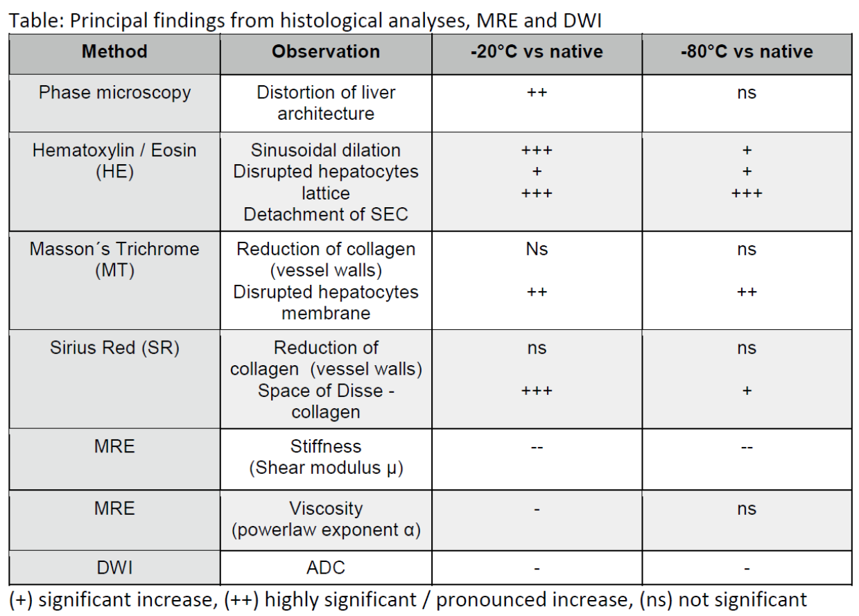
The main histological effects of freezing procedures on tissue structures were identified as follows: i) distortion of liver stroma architecture (visible by phase-microscopy at larger scales in -20°C-tissue, Fig.2), ii) detachment of sinusoidal endothelial cells, SEC (seen by nuclei of SEC which appear rounded in the space of Disse[9], Fig.2), iii) sinusoidal dilatation (apparent by larger sinusoidal sizes at -20°C than -80°C, Fig.2), iv) disruption of hepatocytes membranes (analyzed by the differential uptake of MT dyes for both freezing temperatures, not-shown), v) reduction/modification of sinusoidal collagen (Fig.3). Overall, -80°C-frozen/thawed livers showed better preserved tissue structures than -20°C-frozen/thawed samples. However, both procedures induced significant membrane disruption of hepatocytes, detachment of SEC and collagen disruption at the space of Disse. Fig.4 shows group-values obtained by MRE and DWI. A pronounced decrease of μ due to freezing was measured by MRE while α changed only at -20°C. ADC also reduced significantly after freezing but was not different between the two freezing procedures. Fig.5 summarizes the main findings of this study.Discussion
Since DWI is sensitive to intracellular water mobility, the observed reduction of ADC alludes to distortions of cellular structures leading to hindered water diffusion. The paralleled reduction of stiffness demonstrates the relevance of cellular integrity for the liver’s macroscopic mechanical support. We noted that cells remained at place after freezing/thawing indicating that the geometry of the viscoelastic lattice was less affected by freezing than lattice-inherent rigidity. Probably for that reason α, which is known to be sensitive to the geometrical architecture of tissues[7], remained unchanged by freezing at -80°C which induced fewer structural distortions than ‑-20°C. These distortions were identified by reduction of collagen at the space of Disse, which seems to support the cellular lattice between made of hepatocytes, SEC and Kupffer cells[10]. In summary, deterioration of cell membranes, detachment of SEC and loss of sinusoidal collagen after freezing/thawing reduced the macroscopic shear modulus of liver tissue and affected tissue viscosity. Furthermore, we inferred from histological analysis and MRE-viscosity measurements that collagen structures at the space of Disse are better preserved at -80°C than -20°C.Acknowledgements
Support of the German Research Foundation (GRK2260, BIOQIC) is gratefully acknowledged.References
1. Perepelyuk M, Chin L, Cao X, van Oosten A, Shenoy VB, Janmey PA, Wells RG. Normal and Fibrotic Rat Livers Demonstrate Shear Strain Softening and Compression Stiffening: A Model for Soft Tissue Mechanics. PLoS One 2017;11(1):e0146588.
2. Le Bihan D. Apparent diffusion coefficient and beyond: what diffusion MR imaging can tell us about tissue structure. Radiology 2013;268(2):318-322.
3. Hirsch S, Braun J, Sack I. Magnetic Resonance Elastography: Physical Background And Medical Applications: Wiley-VCH; 2017.
4. Lu YC, Kemper AR, Untaroiu CD. Effect of storage on tensile material properties of bovine liver. J Mech Behav Biomed Mater 2014;29:339-349.
5. Ayyildiz M, Aktas RG, Basdogan C. Effect of solution and post-mortem time on mechanical and histological properties of liver during cold preservation. Biorheology 2014;51(1):47-70.
6. Braun J, Tzschatzsch H, Korting C, Ariza de Schellenberger A, Jenderka M, Driessle T, Ledwig M, Sack I. A compact 0.5 T MR elastography device and its application for studying viscoelasticity changes in biological tissues during progressive formalin fixation. Magn Reson Med 2017:DOI: 10.1002/mrm.26659.
7. Sack I, Joehrens K, Wuerfel E, Braun J. Structure sensitive elastography: on the viscoelastic powerlaw behavior of in vivo human tissue in health and disease. Soft Matter 2013;9(24):5672 - 5680.
8. Reese TG, Heid O, Weisskoff RM, Wedeen VJ. Reduction of eddy-current-induced distortion in diffusion MRI using a twice-refocused spin echo. Magn Reson Med 2003;49(1):177-182.
9. Ishine N, Rubinsky B, Lee CY. A histological analysis of liver injury in freezing storage. Cryobiology 1999;39(3):271-277.
10. Natarajan V, Harris EN, Kidambi S. SECs (Sinusoidal Endothelial Cells), Liver Microenvironment, and Fibrosis. Biomed Res Int 2017;2017:4097205.
Figures