3959
Real-time magnetic resonance elastography: application for monitoring lower leg muscle function during plantar flexion1Charité Universitätsmedizin Berlin, Berlin, Germany
Synopsis
Real-time elastography is well established in ultrasound but has not yet been achieved in MRI. Real-time elastography would be desirable for studying processes that are not easily repeatable or reproducible such as skeletal muscle function. Therefore, we developed a new concept in magnetic resonance elastography (MRE) based on stroboscopic wave sampling and single-shot spiral-k-space acquisition to push temporal resolution of MRE towards real-time elastography. Application of the new method is demonstrated in the lower leg muscles of healthy volunteers.
Introduction
Magnetic resonance elastography (MRE) is an established method for liver fibrosis detection [1] or brain viscoelasticity measurements [2]. However, MRE requires measure times, which make the method susceptible to breathing artefact and prevent direct observation of physiological processes such as muscle function [3]. Therefore, muscle viscoelasticity measurements are mainly done by sonoelastography, which is capable to monitor muscle stiffness changes due to muscle function in real time [4,5]. In this study, we propose real-time MRE based on stroboscopic wave sampling and single-shot spiral-k-space acquisition for direct measurement of skeletal muscle activity.Methods
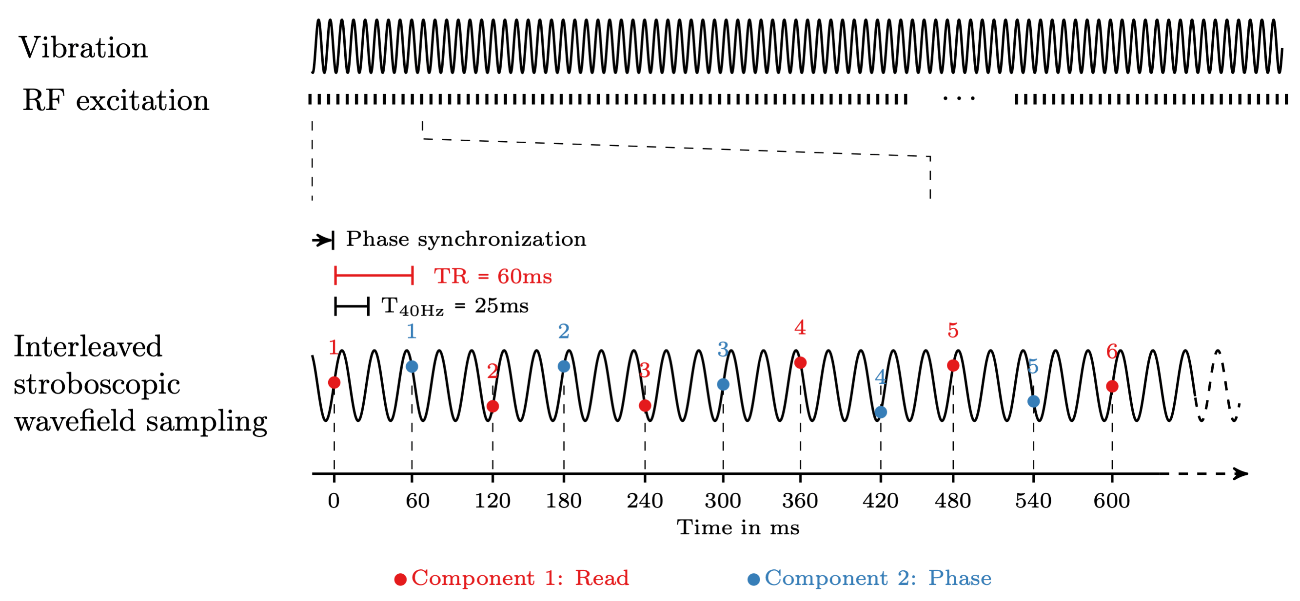
All MRE experiments were performed on a 1.5-T clinical MRI scanner (Magnetom Sonata, Siemens, Erlangen Germany) using a circularly polarized extremity coil. A pressurized air driver was placed below the Achilles’ tendon to inject continuous harmonic vibrations at a frequency of 40 Hz into the lower leg. Figure 1 shows a timing diagram of the experiment. A steady-state MRE image sequence with single-shot spiral readout and stroboscopic wave sampling was used to acquire axial images in the calf, capturing 10 seconds with a frame rate of 16.7 Hz (TR 60 ms). A first order flow-compensated motion-encoding gradient with 30 mT/m amplitude was used (fractional encoding). The echo time was 15 ms, followed by a 40 ms spiral readout and spoiling. We intentionally mismatched the TR and the vibration period to capture different dynamic phases of the wave in a stroboscopic fashion. The motion encoding was only performed in readout and phase encoding direction. This was done in an interleaved fashion, i.e. the odd and even phases were encoded in different directions respectively. Spatial resolution was 1x1x5 mm³. The subject was instructed to wait for 3 seconds after the start of acquisition and then to stretch the foot against the resistance of a rubber foam block that was placed below the sole of the foot. Already during image reconstruction, the first image was used for background phase correction for all other phases. When using multi-channel receive coils, this phase correction results in superior SNR in the combined phase images. MRE postprocessing was based on tomoelastography, adapted to the stroboscopic wavefield sampling approach and provided 82 shear-wave speed (SWS) maps as the surrogate of stiffness in units m/s. Regions of interest (ROI) were drawn in the gastrocnemius, the tibialis anterior and soleus muscles. Stiffness changes over time were compared in these ROIs.Results
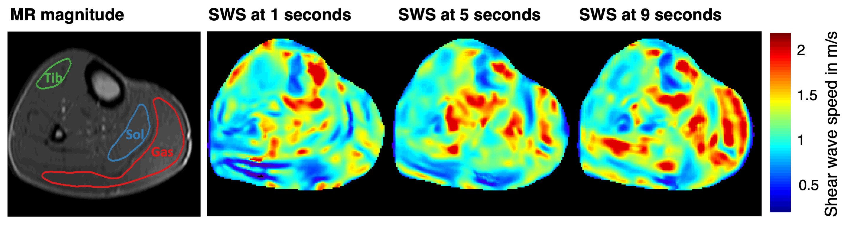
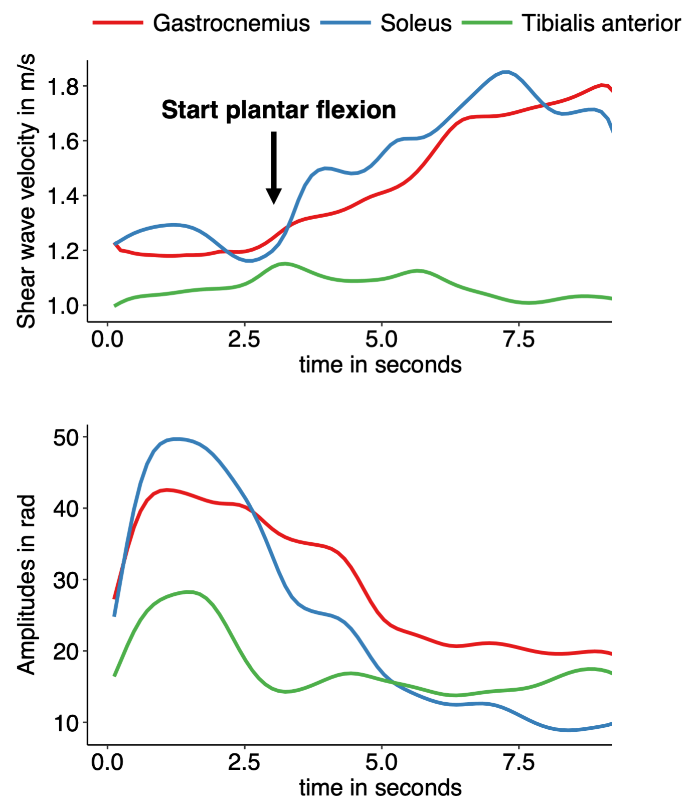
Figure 2 shows 3 of the 82 dynamic elastograms at rest, shortly after the beginning of plantar flexion and 4 seconds later. The time-course of shear wave speed in different muscles of the calf is depicted in figure 3. In the gastrocnemius and soleus, which are primarily responsible for plantar flexion, the shear wave speed increases by about 50% during the task. In contrast, almost no change is detected in the tibialis anterior responsible for flexion in opposite direction. In all ROIs we detected decreased wave amplitudes during flexion. This could indicate a disturbed mechanical coupling of the actuator to the leg caused by the leg movement.Discussion
Our real-time MRE image acquisition is based on steady-state single-shot k-space acquisitions, similar to the concept of real-time MRI of Frahm and colleagues [6], however, without nonlinear inversion reconstructions of highly undersampled MRI datasets. Without the use of parallel imaging techniques, our method is currently limited to relatively small FoVs but extension to this acquisition and reconstruction is planned. Use of stroboscopic sampling of harmonic shear vibrations is well established in time-harmonic ultrasound elastography [7]. We exploited this principle for the first time in MRE. Temporal resolution was significantly improved by neglecting the through-plane wave deflection component. This is justified when observing wave propagation in a transverse view relative to the muscle fibers giving us the opportunity to compute the in-plane curl-component which relates to the isotropic shear modulus μ12 in transversal-isotropic media [8]. More in-depth analysis of this isotropic shear modulus and its variation during muscle activity is required.Conclusion
Real-time MRE is possible, currently in a small FOV with an effective frame rate of 8-9 Hz reconstructed in a sliding-window fashion from adjacent stroboscopic wave imagesAcknowledgements
We acknowledge continuing support by GRK2260 (BIOQIC) of the German Research Foundation and SFB1340.References
1. Venkatesh SK, Yin M, Ehman RL. Magnetic resonance elastography of liver: Technique, analysis, and clinical applications. J Magn Reson Imaging 2013;37(3):544-555.
2. Hiscox LV, Johnson CL, Barnhill E, McGarry MD, Huston J, van Beek EJ, Starr JM, Roberts N. Magnetic resonance elastography (MRE) of the human brain: technique, findings and clinical applications. Phys Med Biol 2016;61(24):R401-R437.
3. Hirsch S, Braun J, Sack I. Magnetic Resonance Elastography: Physical Background And Medical Applications: Wiley-VCH; 2017.
4. Gennisson JL, Deffieux T, Mace E, Montaldo G, Fink M, Tanter M. Viscoelastic and Anisotropic Mechanical Properties of in Vivo Muscle Tissue Assessed by Supersonic Shear Imaging. Ultrasound in Medicine and Biology 2010;36(5):789-801.
5. Deffieux T, Montaldo G, Tanter M, Fink M. Shear Wave Spectroscopy for In Vivo Quantification of Human Soft Tissues Visco-Elasticity. Ieee Transactions on Medical Imaging 2009;28(3):313-322.
6. Uecker M, Zhang S, Voit D, Karaus A, Merboldt KD, Frahm J. Real-time MRI at a resolution of 20 ms. NMR Biomed 2010;23(8):986-994.
7. Tzschatzsch H, Nguyen Trong M, Scheuermann T, Ipek-Ugay S, Fischer T, Schultz M, Braun J, Sack I. Two-Dimensional Time-Harmonic Elastography of the Human Liver and Spleen. Ultrasound Med Biol 2016;42(11):2562-2571.
8. Guo J, Hirsch S, Scheel M, Braun J, Sack I. Three-parameter shear wave inversion in MR elastography of incompressible transverse isotropic media: Application to in vivo lower leg muscles. Magn Reson Med 2016;75(4):1537-1545.
Figures