3958
Characterization of fibril aggregates in ex vivo rat brain with multi-frequency MR Elastography – Preliminary results1Univ. Lyon, INSA-Lyon, Université Claude Bernard Lyon 1, UJM-Saint Etienne, CNRS, Inserm, CREATIS UMR 5220, U1206, Lyon, France, 2Univ. Lyon, Lyon Neuroscience Research Center, CNRS UMR 5292, INSERM U1028, Univ. Lyon 1, Lyon, France, 3INSERM U 1148, Laboratory of Vascular Translational Science; X. Bichat Hospital University Paris Diderot, Paris, France
Synopsis
Fibrils are biomarkers for early stages of dementia. In this work, α-synuclein fibrils were injected in rat striatum. Brains were imaged ex-vivo using multi-frequency MR Elastography. Estimation of the real part kr of the complex wave number was made for each studied frequency in ROIs surrounding the inclusion, a contralateral control injection and the whole brain. Exponent of the frequency power law was derived from kr maps acquired at different frequencies. No difference was observed between kr values for the different ROIs, but the exponent was more important at the fibrils location, potentially indicating that multi-frequency MRE can detect fibrils.
Introduction
Accurate and early diagnosis of dementia represents a major challenge in an ageing society. Pathological hallmarks of these diseases are fibrils, formed by the aggregation of endogenous proteins misshaped to β-sheet conformation. Accurate localization of these fibrils could improve diagnosis given that diverse dementias protein accumulation starts in different locations.
We suggest using multi-frequency Magnetic Resonance Elastography (MRE) to characterize those aggregates. MRE has already been used to characterize the spatial distribution of sub-voxel obstacles1,2. Other groups have found stiffness changes in transgenic mice3–5; however the substrate of these changes is not well characterized: effects of fibrils, neuronal loss, vascularization or inflammation are not discriminated. The purpose of this work is to characterize the local changes induced by injected fibrils on the brain mechanical properties
Methods
Four Sprague Dawley rats were injected with 10µL of α-synuclein fibrils at 200µM in the striatum area. The opposite hemisphere received a control injection of Phosphate Buffered Saline (PBS). Animals were sacrificed 2 to 4 weeks after injections. The brains were immediately embedded in 1% agarose gel (SIGMAF8630) with artificial cerebrospinal fluid6. The brain was then imaged on a Bruker 4.7T scanner with a home-designed volume coil dedicated to small sample MRE. RARE sequence was used as anatomical image. MRE measurements were performed at 800, 900, 1000 and 1200Hz. 1000Hz acquisition was done first and repeated before the last frequency was acquired, to confirm that brain mechanical properties were not degraded during acquisition time (90 minutes). MRE sequence was derived from RARE sequence7. Motion was encoded in the three directions with the following parameters: 8 snapshot of the waves, 7 1-mm-thick slices, FOV of 2.5x2.5cm², in plane matrix of 64×64, leading to voxel size of 0.391x0.391x1mm3.
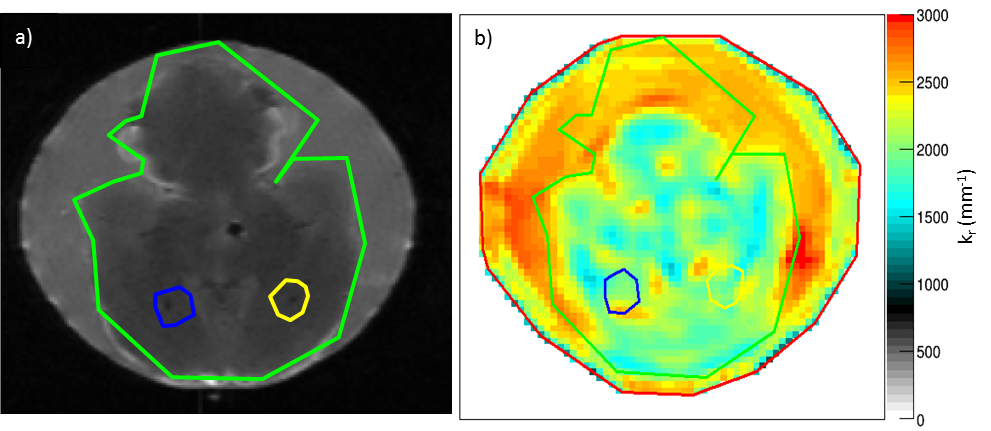
Processing involves a 3D direct curl inversion8. Reconstruction was performed with the application of a Gaussian filter (σ = 0.39x0.39x1mm3, kernel = 3x3x3 voxels) on the unwrapped phase images. Real part kr (inversely proportional to wave speed) of the complex wave number was estimated in manually defined ROIs corresponding to the whole brain, the α-synuclein injection localization and the contralateral injection. Those ROIs are reported on anatomical and kr reconstructed images (Fig.1). The exponent y of the frequency power law demonstrated by Lambert et al.2 was derived from kr maps reconstructed at different frequencies for each ROI. Allometric fitting (a*frequencyy) was realized using Origin software (OriginPro 2016 64-bit, http://www.OriginLab.com). Paired sample Wilcoxon tests were performed to compare the dispersion in the ROIs.
Results
Mechanical parameters of the brain remained stable during whole imaging duration: for the two 1000Hz acquisitions, reconstructed kr varied in average by 9 ± 6% for the whole brain, 13 ± 9% for the fibril injection and 4 ± 3% for the PBS injection. For the whole brain ROI, this variation is inferior to the standard deviations of each kr measured at 1000 Hz.
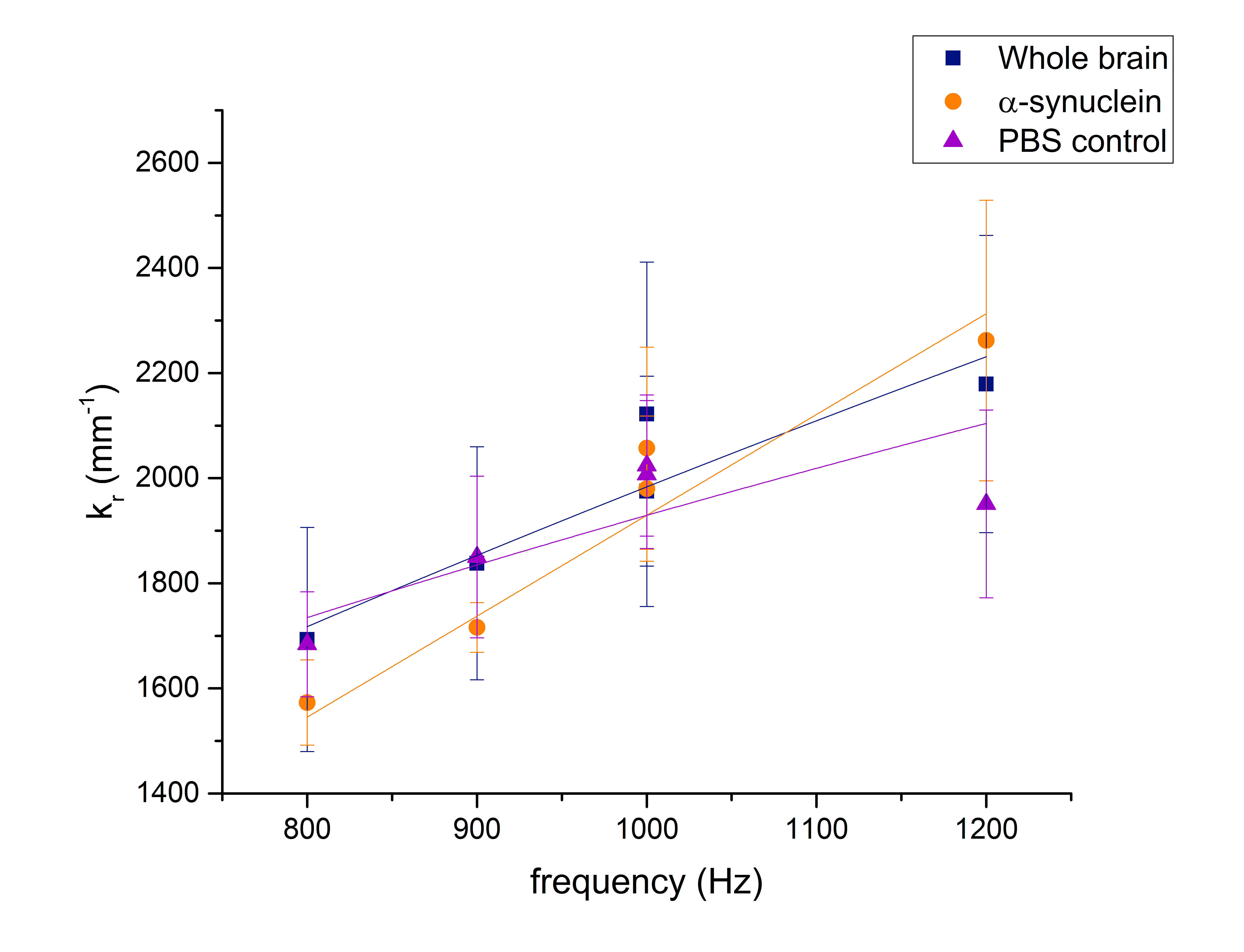
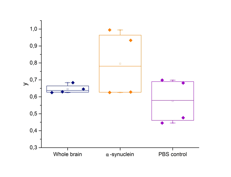
No significant difference was found between kr estimated from the three ROIs whatever the frequency investigated (Fig.2). Interestingly, y was superior in average in the α-synuclein ROI (0.80 ± 0.09) compared to both the whole brain (0.65 ± 0.07) and the contralateral PBS injection (0.58 ± 0.04) (Fig.3). In two cases, y in the injection ROI was close to y in the whole brain. For two rats, y in PBS seemed close to y in the whole brain, but less for two others. Wilcoxon tests did not qualify this augmentation as significant.
Discussion and conclusions
Single-frequency MRE for fibril aggregates detection does not appear relevant as no change in kr was detected whatever the frequency investigated. These results support recent findings in Alzheimer’s transgenic animals suggesting that fibrils alone are not responsible for viscoelasticity changes at 1000Hz3.
This work shows for the first time that α-synuclein fibrils, injected in an important quantity without over toxicity and inflammation at the sub-acute stage, can hardly be detected using multi-frequency MRE. An increase of the parameter y is observed at the fibril location. This parameter could be sensitive to microarchitecture alteration, as indicated by Schregel et al.9. A decrease of this same parameter is observed in a contralateral PBS injection. These changes are not significant, due to the small number of imaged animals, but also to some values, very close to whole brain dispersion. These values could be explained by a wrong estimation of the injection area when placing the ROIs, which will need validation from ongoing histological analysis.
Additional acquisitions are needed to confirm these results. The sensitivity of the method to detect the presence of fibrils must be studied by decreasing the concentration of injected fibrils.
Acknowledgements
This work was supported by the LABEX PRIMES (ANR-11-LABX-0063) of Université de Lyon, within the program "Investissements d'Avenir" (ANR-11-IDEX-0007) operated by the French National Research Agency (ANR) and by PEPS CNRS “Balanced”. We want to acknowledge the PILoT imaging platform, member of France Life Imaging network (grant ANR-11-INBS-0006).for the support provided on image acquisition.References
1. Jugé L, Petiet A, Lambert SA, et al.: Microvasculature alters the dispersion properties of shear waves--a multi-frequency MR elastography study. NMR Biomed 2015; 28:1763–1771.
2. Lambert SA, Näsholm SP, Nordsletten D, et al.: Bridging Three Orders of Magnitude: Multiple Scattered Waves Sense Fractal Microscopic Structures via Dispersion. Phys Rev Lett 2015; 115:094301.
3. Majumdar S, Mishra R, Lazarov O, Klatt D: Early-stage analysis of murine models of Familial Alzheimer’s disease: Preliminary results. 1st MRE Workshop Berlin, Germany; 2017:25.
4. Murphy MC, Curran GL, Glaser KJ, et al.: Magnetic resonance elastography of the brain in a mouse model of Alzheimer’s disease: initial results. Magn Reson Imaging 2012; 30:535–539.
5. Munder T, Pfeffer A, Schreyer S, et al.: MR elastography detection of early viscoelastic response of the murine hippocampus to amyloid β accumulation and neuronal cell loss due to Alzheimer’s disease. J Magn Reson Imaging 2017; 47:234–245.
6. Preparation of Artificial CSF [http://www.alzet.com/products/guide_to_use/cfs_preparation.html]
7. lefebvre P, Tse Ve Koon K, Brusseau E, et al.: Comparison of viscoelastic property characterization of plastisol phantoms with magnetic resonance elastography and high-frequency rheometry. EMBC Orlando, United States; 2016.
8. Sinkus R, Daire J-L, Beers BEV, Vilgrain V: Elasticity reconstruction: Beyond the assumption of local homogeneity. C R Mécanique 2010; 338.
9. Schregel K, Wuerfel E, Garteiser P, et al.: Demyelination reduces brain parenchymal stiffness quantified in vivo by magnetic resonance elastography. Proc Natl Acad Sci U S A 2012; 109:6650–6655.
Figures