3957
In vivo Assessment of Gray Matter Stiffness and Perfusion in the Human Brain1Department for Diagnostic Physics, Oslo University Hospital, Oslo, Norway, 2The Intervention Center, Oslo University Hospital, Oslo, Norway, 3Division of Imaging Sciences & Biomedical Engineering, King's College London, London, United Kingdom, 4Inserm U1148, LVTS, University Paris Diderot, Paris, France
Synopsis
MR Elastography (MRE) is an emerging imaging technique used to quantify the stiffness and viscosity of tissue. Few studies have investigated the relationship between these biomechanical properties and brain perfusion. If understood, this could provide valuable new insight in diseases that alter biomechanical properties or functionality. In our study we assess the cerebral blood flow and the tissue stiffness of gray matter in healthy volunteers. We find that in cortical gray matter, both the cerebral blood flow and the shear storage modulus G’ was higher than in deep gray matter.
Introduction
Methods
This preliminary study was performed in six healthy volunteers, and approved by the Regional Ethics Committee, including the use of informed consent. The exams were performed on a 3T MRI scanner (Philips Ingenia), and the MR Elastography was performed using the Gravitational Transducer2, which induced shear waves of 50 Hz into the head of the volunteers. Full wave field data were acquired in 19 continguous transversal slices of 3.06×3.14×3.14 mm resolution with a GR-EPI readout. Further imaging parameters were: Hadamard3 encoding scheme, echo time TE =11.51 ms, repetition time TR=380 ms, matrix size 72×70, motion encoding gradient strength of 19.9/34.6 mT/m, and five acquisitions over a wave cycle. The MRE scan time was 4.5 minutes.
To estimate brain perfusion, we performed an Arterial Spin Labelling (ASL), 2D acquisition with a labelling duration and a post labelling delay of 1.8 s. We acquired 35 pairs of label and control volumes, with echo time TE =14.8 ms, repetition time TR=440 ms, and a resolution of 3.6×3.6×5.5 mm.
Image analysis of the ASL images was done in Nordic
ICE (Nordic Neuro Lab, version 1.0.0.0), and the elastography images were
analyzed using an established approach4. Regions-of-interest (ROIs)
were placed manually in caudate nucleus (deep gray matter) and Broca’s area (cortical
gray matter). Two ROIs were measured for each scan, and the MRE values
were also averaged over two scans.
Results
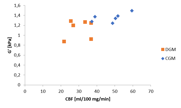
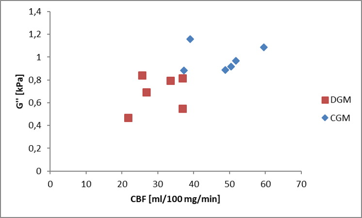
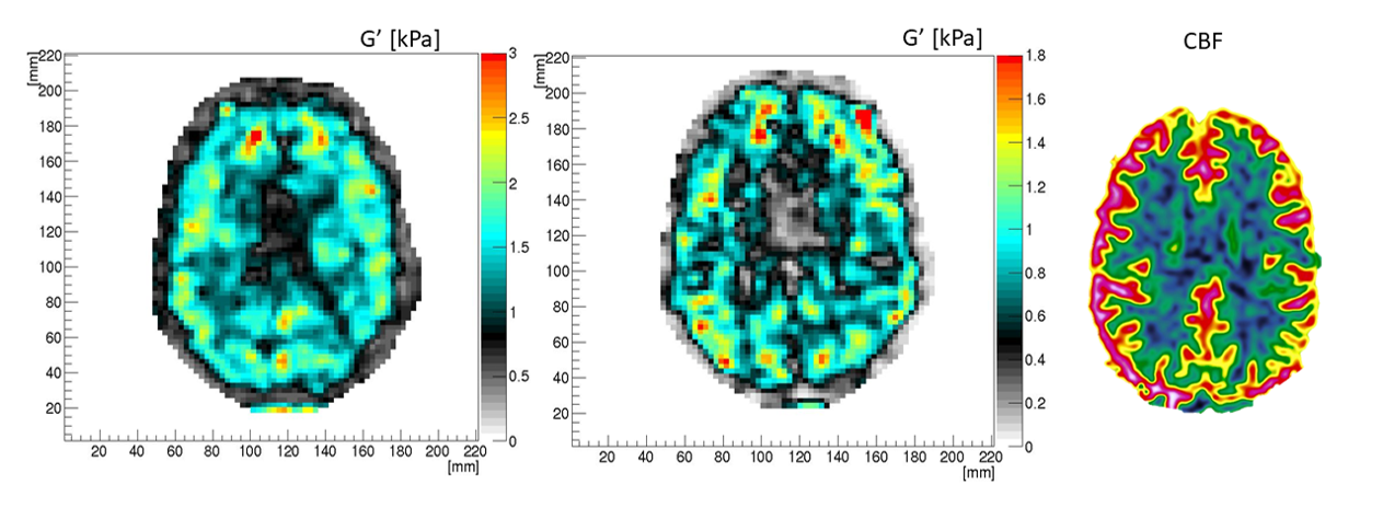
As shown in Figure 1, the cerebral blood flow (CBF) is higher in the cortical gray matter than in the deep gray matter, and the shear storage modulus G’ also shows a tendency towards higher values in cortical gray matter. For the shear loss modulus G’’, the values for both the shear loss modulus and the perfusion are higher in cortical gray matter, shown in Figure 2. Comparing the values for cortical and deep gray matter related to CBF for both G’ and G’’, a Wilcoxin test yields differences of p<0.05. Figure 3 shows example images of G’, G’’ and CBF for one of the volunteers.Discussion
In this preliminary work, we found that in cortical gray matter, both the cerebral blood flow and the shear storage modulus G’ was higher than in deep gray matter. Results recently published3 on ASL and MRE in sub-regions of deep gray matter, suggested a high sensitivity of MRE in deep gray matter to perfusion pressure. The relationship between the two is likely to be affected by tissue and vascularity changes in diseases such as cancer, and further studies in patients is required to learn about how these properties will affect each other.Conclusion
Further work is needed to understand the relationship between biomechanical properties of brain tissue and vascularity, in both the healthy and patient population. In this limited sample, we show that both the perfusion and stiffness were higher in the cortical gray matter than in the deep gray matter.Acknowledgements
No acknowledgement found.References
1. Muthupillai R, Lomas DJ, Rossman PJ, et al. Magnetic resonance elastography by direct visualization of propagating acoustic strain waves. Science. 1995;269(5232):1854-1857
2 . Runge JH, Hoelz SH, Sudakova J, et al. A novel MR Elastography transducer concept based on a rotational eccentric mass: the gravitational transducer. ISMRM. Honolulu 2017
3. Guenthner C, Runge JH, Sinkus R et al. Analysis and improvement of motion encoding in magnetic resonance elastography. NMR in Biomedicine.2018;31:e3908.
4. Sinkus R, Tanter M, Xydeas T, el al. Viscoelastic shear properties of in vivo breast lesions measured by MR elastography. Magn Reson Imaging. 2005;23(2 SPEC. ISS.):159-165.).
5. Hetzer S, Birr P, Fehlner A, et al. Perfusion alters stiffness of deep gray matter. Journal of Cerebral Blood Flow & Metabolism. 2018;38(1):116–125
Figures