3955
Multi-Excitation MRE in Aging Human Brain: Estimation of Direction-Dependent Mechanical Properties1Biomedical Engineering Department, Illinois Institute of Technology, Chicago, IL, United States, 2Beckman Institute, University of Illinois at Urbana-Champaign, Champaign, IL, United States, 3Department of Mechanical & Aerospace Engineering, Rutgers University, Piscataway, NJ, United States
Synopsis
Healthy aging affects the local mechanical properties of the human brain tissue but their estimation remains a challenge, especially in strongly anisotropic white matter regions. This study merges previous MRE aging research involving high-resolution, full-coverage, and multi-excitation MRE imaging with the ability to estimate anisotropic shear moduli locally. We identified important anisotropic differences in the storage modulus in the corpus callosum, which help differentiate old from the young.
INTRODUCTION
Using isotropic tissue models for inversion, prior MRE studies have shown that there is a general decrease of the human brain stiffness during normal aging [1-5]. A class of anisotropic inversion methods rely on directional filtering of the displacement fields in judiciously chosen regions of the brain, followed by direct extraction of certain components of the stiffness tensor, which are assumed uniform in the chosen regions [6,7]. Romano et al. [6] found that one of the shear moduli decreases in the corticospinal tract of amyotrophic lateral sclerosis patients in-vivo. Schmidt et al. [7] reported good agreement of the anisotropic moduli with shear tests in an ex-vivo study of the porcine corpus callosum. Here we report a novel iterative inversion scheme based on a transversely isotropic tissue model, and we apply it for the in-vivo study of the human corpus callosum. We incorporate the data obtained in multi-excitation MRE and diffusion tensor imaging (DTI) study of the brain [8,9] in an iterative scheme that allows the estimation of shear moduli parallel and perpendicular to the local axon direction, and then compare the regional average values between two cohorts of healthy young and old adults.
METHODS
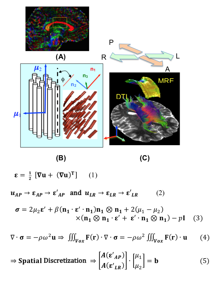
A previous study [9] generated anterior-posterior (AP) excitation MRE, and left-right (LR) excitation brain MRE data for four young (24-32 years old) and 4 older (55-76 years old) males. Using a 3D multislab, multishot spiral MRE sequence [10], 3D complex displacement field data was collected at 50 Hz with 2mm3 isotropic spatial resolution. The isotropic viscoelastic properties were estimated for both AP and LR data using nonlinear inversion (NLI). Additionally, matched field-of-view and resolution (relative to MRE) diffusion tensor imaging (DTI) and high-resolution anatomical MPRAGE at 0.9mm3 isotropic resolution (TR/TI/TE = 2000/900/2.2 ms) were acquired, to register the subjects to the MNI JHU white matter atlas. Fig. 1 presents the scheme introduced here, which is labeled as ITI, for “Inverse Transversely Isotropic”. Under ITI, we use both AP and LR data, in conjunction with DTI, to extract the shear moduli of the transversely isotropic model. Fig. 1(A), focuses on an ROI consisting of 18x18x18 voxels, centered on the body of the corpus callosum of each subject. Based on Eq. (1), strain tensors for AP and LR excitations are obtained from the 3D displacement fields, as measured by MRE on world coordinates. For each voxel the strain fields are transformed to the local coordinate system aligned with the principal fiber direction Fig. 1(B). The linear stress-strain relationship for a nearly-incompressible, transversely isotropic medium is given by Eq. (3). The shear moduli correspond to shear stiffness parallel (μ1) and transverse (μ2) to the principal fiber direction, both complex in the case of harmonic waves in viscoelastic materials. Here we take the ratio of E1 / E2 = 2.7 [11] and ν12 = 0.4999. ITI inversion involves the following steps: a weak form of the governing equation is derived by multiplying it with a judiciously chosen function F(r) and integrating over the voxel volume(Eq. (4)). The integral is discretized using a higher order spatial scheme, which results in a system of linear equations for the pair of complex moduli that is solved iteratively.
RESULTS AND DISCUSSION
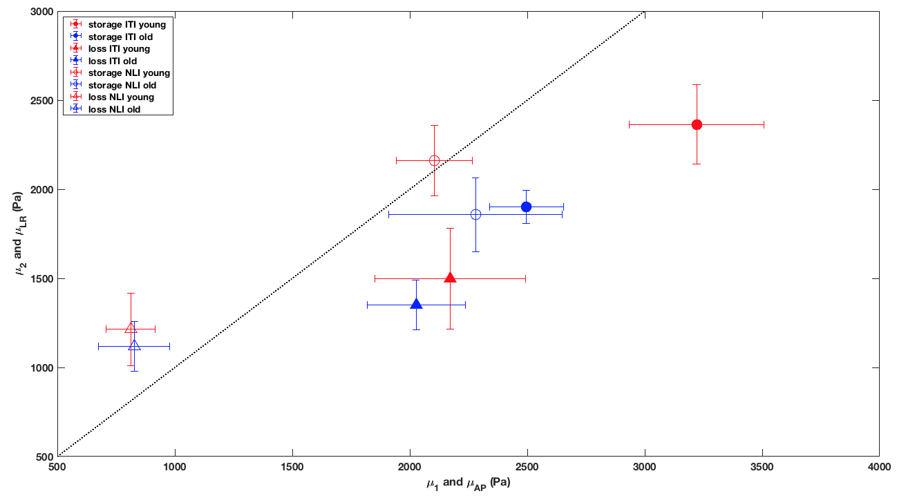
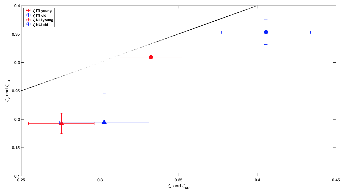
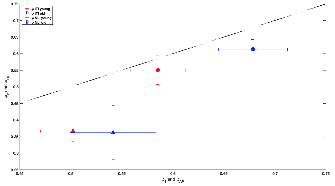
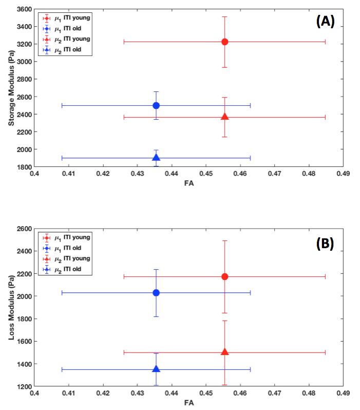
Fig. 2 indicates that the anisotropic model (ITI) accentuates the difference between young and old groups in terms of shear moduli, relative to NLI. Both models show decrease of storage moduli with age, but the ITI shows decrease of both storage and loss moduli. Both models show a higher damping ratio and phase angle for the old group (Fig. 3 and 4), where the ITI model enhances those differences. Fig. 5 shows the correlation of the fractional anisotropy with the loss and storage moduli obtained by ITI. The model show and increase in modulus with increasing FA for both groups.CONCLUSION
The work presented here demonstrates the benefit in mechanical properties reconstruction by shaking a human head in two different directions (AP and LR). A novel iterative (Inverse Transversely Isotropic, ITI) scheme, incorporating pairs of AP and LR data sets and DTI, allowed the estimation of anisotropic shear moduli in the corpus callosum of a young and an older group. This study also underlines the importance of accurately modeling certain brain white matter regions by anisotropic tissue models.Acknowledgements
Support was provided by NSF Grants CMMI-1437113 and CMMI-1762774. Partial support was provided by the Biomedical Imaging Center of the Beckman Institute for Advanced Science and Technology at the University of Illinois at Urbana-Champaign (UIUC), NIH Grant R01-EB018230, and NIH/NIBIB Grant R01-EB001981.References
[1] Sack, I, Beierbach, B, et al. The impact of aging and gender on brain viscoelasticity. NeuroImage. 2009;46, 652–657.
[2] Sack, I, Streitberger, KJ, et al. The Influence of Physiological Aging and Atrophy on Brain Viscoelastic Properties in Humans. PLoS ONE. 2011;6, e23451.
[3] Guo, J, Hirsch, S, et al. Towards an Elastographic Atlas of Brain Anatomy. PLoS ONE. 2013;8, e71807.[4] Arani, A, Murphy, MC, et al. Measuring the effects of aging and sex on regional brain stiffness with MR elastography in healthy older adults. NeuroImage. 2015;111, 59–64.
[5] Murphy, MC, Huston J, et al.. MR elastography of the brain and its application in neurological diseases. Neuroimage 2018, in press.
[6] Romano A, Guo J, et al. In vivo waveguide elastography: Effects of neurodegeneration in patients with amyotrophic lateral sclerosis. Magn. Reson. Med. 2014;72(6):1755-1761.
[7] Schmidt JL, Tweten DJ, et al. Measurement of anisotropic mechanical properties in porcine brain white matter ex vivo using magnetic resonance elastography. JMBBM. 2018;79:30-37.
[8] Anderson, AT, Van Houten, EEW, et al. Observation of direction-dependent mechanical properties in the human brain with multi-excitation MR elastography. JMBBM. 2016;59, 538–546.
[9] Anderson, AT, Johnson, CL, et al., Multi-Excitation MRE in Aging Human Brain. 26th Annual Meeting of ISMRM. 2018.
[10] Johnson, CL, Holtrop, JL, et al. 3D multislab, multishot acquisition for fast, whole-brain MR elastography with high signal-to-noise efficiency. Magn. Reson. Med. 2014; 71, 477–485.
[11] Feng Y, Okamoto RJ, et al. Measurements of mechanical anisotropy in brain tissue and implications for transversely isotropic material models of white matter. JMBBM. 2013;23:117–132.
Figures