3948
SSFP-fMRI: Radial reading helps to Improve Temporal SNR1School of Cognitive Science, Institute for Research in Fundamental Sciences (IPM), Tehran, Iran (Islamic Republic of), 2Department of Biomedical Engineering, Amirkabir University of Technology, Tehran, Iran (Islamic Republic of)
Synopsis
SSFP allows us to have functional contrast close to spin-echo, but it suffers from low temporal-resolution. Increasing
Introduction
Steady-State Free Precession (SSFP)-fMRI is an attractive technique since it obtains clean functional contrast (close to spin-echo) for high-spatial mapping of the neural activity1, 2, 3. In this technique, echoes are collected between repetitive RF pulses with short TRs. SSFP-fMRI, therefore, has low temporal resolution that results in limited spatial coverage4. Temporal SNR (tSNR) enhancement is a remedy for this limitation as it decreases the number of required volumes for determining activities with a specific confidence. For SSFP, in contrast to GRE-EPI, the Radial acquisition is quite straightforward. Since the dense sampling of the central part of k-space, where the energy of the signal is mostly concentrated, leads to reduce noise, motion artifacts and off-resonance effects5. We hypothesize that such an acquisition in SSFP-fMRI can improve its tSNR and consequently decrease the acquisition time. Here we test this hypothesis along with the robustness of Radial acquisition of SSFP for fMRI.Method
Data acquisition: 5 healthy subjects were recruited and scanned at a 3T MRI scanner (Siemens, Trio Tim). For each subject, two sets of data were acquired based on 2D balanced SSFP sequence with 50 preparation pulses for Cartesian and Radial k-space trajectories using a 12-channel head coil with identical imaging parameters as follows: TR/TE = 6.12/3.06 ms, squared FOV = 224 mm, number of Phase-Enc. /Spokes = 112, Flip Angle = 30°, Voxel size = 2×2×3 mm3 and 72 measurements. Radial acquisitions were reconstructed by the routine re-gridding method, available on the scanner. Due to limited coverage, only 4 coronal slices were selected in the occipital lobe, which is specific for the visual stimulus task. The task consisted of 6 blocks (24s-24s) with the volume TR of 4s, which at the act-state, a rotating checkerboard was presented on back-projected screen.
Data analysis pipeline: All the preprocessing and analysis steps were performed using FSL (FMRIB, Oxford Univ.). Motion correction, temporal filtering (cut off frequency= 120s) and spatial smoothing (FWHM=4mm) were applied in all cases as a preprocessing step. For statistical inference, a general linear model (GLM) was used as implemented in FSL Feat. For generating activation maps cluster-based thresholding with z-score=3 and p=0.05 was applied for both Radial and Cartesian acquisitions. To evaluate the tSNR as a measure of signal stability, the mean of each time series was divided by its standard deviation in rest conditions.
Results
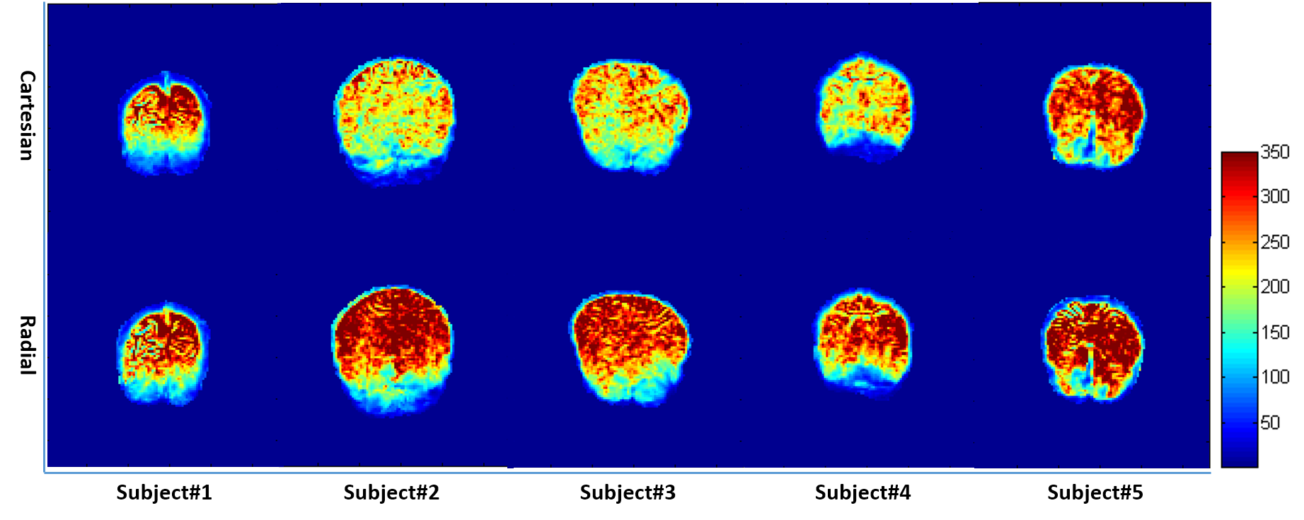
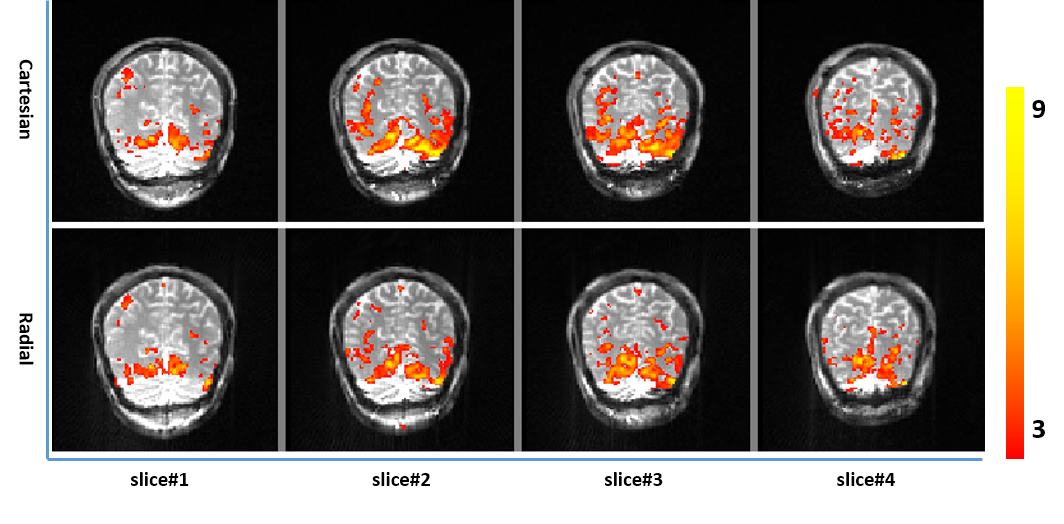
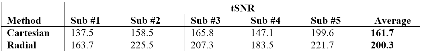
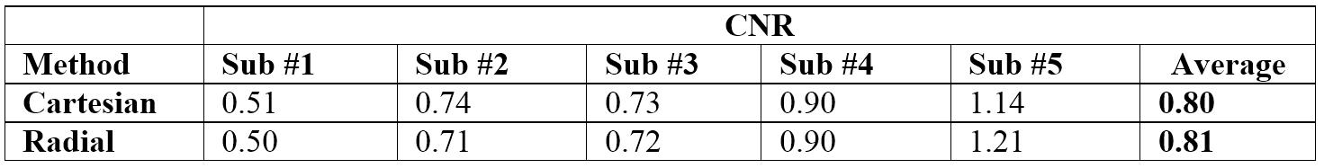
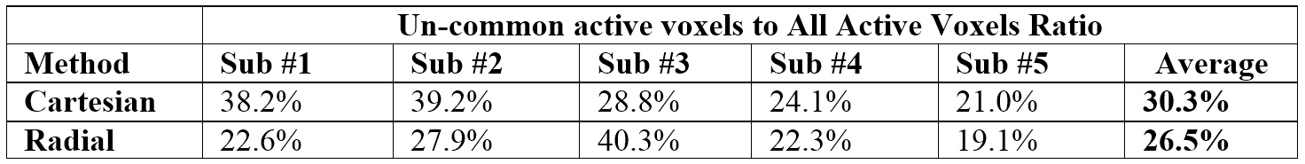
Temporal SNR maps of the Cartesian and Radial acquisitions in coronal view are shown in Figure 1 for a selected slice of each subject. Figure 2 shows the activation maps for a representative subject, displayed for all slices. As seen in this figure, in both techniques, consistent visual activity with similar patterns is indicated in the expected brain regions related to the task. In Table 1 & 2, tSNR and functional CNR values for all subjects were reported on middle slices. The average values for tSNR and CNR are equal to 200.3 & 0.80 for Radial and 161.7 & 0.81 for Cartesian acquisitions, respectively. The percent of active voxels (with a z-score threshold of 3 or more) that were collected only by one technique alone compared to all active voxels were shown in Table 3. The higher the values shown in table 3, the lower the expected specificity of the technique.Discussion and Conclusion
Here, we acquired data with identical imaging parameters, such as the number of k-space lines, TRs, and spatial resolution, for both Cartesian and Radial SSFP approaches. The results confirm that Radial approach has higher tSNR compared to Cartesian one (Table 1 & Fig. 1). This can be occurred due to the dense oversampling in center of K-Space for Radial acquisition and motion robustness of this technique due to the equal contribution of each spoke into image contrast. Moreover, Radial SSFP shows the robust activation with the functional sensitivity close to the Cartesian acquisition (Table 2 & Fig. 2) but with the less un-common active voxels which can be interpreted as a lower false positive rate (Table 3). In conclusion, Radial acquisition is an alternative robust fMRI method with an advantage of higher tSNR compared to Cartesian one that can help to improve the temporal resolution and reducing false-positives activations in SSFP-fMRI applications.Acknowledgements
No acknowledgement found.References
1. Scheffler, K., Seifritz, E., Bilecen, D., Venkatesan, R., Hennig, J., Deimling, M., & Haacke, E. M. (2001). Detection of BOLD changes by means of a frequency‐sensitive trueFISP technique: preliminary results. NMR in Biomedicine: An International Journal Devoted to the Development and Application of Magnetic Resonance In Vivo, 14(7‐8), 490-496.
2. Barth, M., Meyer, H., Kannengiesser, S. A., Polimeni, J. R., Wald, L. L., & Norris, D. G. (2010). T2‐weighted 3D fMRI using S2‐SSFP at 7 tesla. Magnetic Resonance in Medicine: An Official Journal of the International Society for Magnetic Resonance in Medicine, 63(4), 1015-1020.
3. Malekian, V., Moghaddam, A. N., & Khajehim, M. (2018). A robust SSFP technique for fMRI at ultra-high field strengths. Magnetic resonance imaging, 50, 17-25.
4. Lee, J. H., Hargreaves, B. A., Hu, B. S., & Nishimura, D. G. (2003). Fast 3D imaging using variable‐density spiral trajectories with applications to limb perfusion. Magnetic Resonance in Medicine: An Official Journal of the International Society for Magnetic Resonance in Medicine, 50(6), 1276-1285.
5. Lee, J. H., Hargreaves, B. A., Hu, B. S., & Nishimura, D. G. (2003). Fast 3D imaging using variable‐density spiral trajectories with applications to limb perfusion. Magnetic Resonance in Medicine: An Official Journal of the International Society for Magnetic Resonance in Medicine, 50(6), 1276-1285.
Figures