3946
Comparison of continuous sampling with active noise cancellation and sparse sampling for cortical and subcortical auditory fMRI1Sir Peter Mansfield Imaging Centre, School of Physics and Astronomy, University of Nottingham, Nottingham, United Kingdom, 2NIHR Nottingham Biomedical Research Centre, Nottingham, United Kingdom, 3Hearing Sciences, Division of Clinical Neuroscience, School of Medicine, University of Nottingham, Nottingham, United Kingdom, 4University of Nottingham Malaysia, Jalan Broga, Selangor Darul Ehsan, Malaysia, 5Manchester Centre for Audiology and Deafness (ManCAD), University of Manchester, Manchester, United Kingdom, 6NIHR Manchester Biomedical Research Centre, Manchester, United Kingdom, 7Department of Psychology, Lancaster University, Lancaster, United Kingdom
Synopsis
We compared cortical and subcortical auditory BOLD fMRI responses to epochs of broadband noise acquired using continuous sampling with active noise cancellation (TR = 2s) to sparse sampling (TR = 8s, 1993ms acquisition with 6007ms silence) in a repeated measures factorial design. High resolution (1.5 mm3) 3 Tesla fMRI data were acquired using multiband 2 and corrected for physiological noise and image distortions. BOLD beta estimates are compared for onset, offset and sustained auditory fMRI responses for both sampling schemes. We report the strengths and weaknesses of the two sampling methods.
Introduction
fMRI of the auditory pathway requires perception of a salient auditory stimulus compared to a “silent” baseline period. It has been shown that “higher” sections of the pathway (upper-midbrain/cortex) respond preferentially to transient stimuli whilst “lower” regions (brainstem) respond more to sustained acoustic energy [1]. However, the baseline period of auditory fMRI is confounded by acoustic noise from imaging gradients. Two approaches to reduce the impact of scanner acoustic noise are (i) “sparse” temporal sampling of single brain volumes, providing intervals with no scanner acoustic noise [2]; or (ii) active noise cancellation, with commercially-available systems generating acoustic signals that add destructively with the scanner acoustic noise to significantly reduce the overall acoustic energy at the eardrum of the participant [3]. Here, we use a broadband noise block design for optimal activation of subcortical regions, comparing these two sampling approaches to assess sustained and transient auditory responses in subcortical and cortical auditory regions.Methods
15 participants (n=11 female; age=33±11 years) with self-reported normal hearing were scanned on a Philips Ingenia 3.0 T MR scanner with 32-channel head coil using a gradient-echo EPI readout (TE=34ms, FOV=168×168mm, 1.5mm isotropic resolution, SENSE2, multiband2, halfscan=0.927). 46 coronal-oblique slices provide coverage of the brainstem and Heschl’s gyrus. Continuous sampling was performed with a TR=2s and equidistant temporal slice spacing, sparse sampling had a 1993ms image acquisition (TA) followed by 6007ms silence (effective TR=8s).
Each fMRI run comprised 12 cycles of 24-s broadband noise (1.4-4.1 kHz; 85 dB-SPL) and 24-s rest resulting in 144/36 acquisitions for noise and rest for continuous/sparse sampling. Stimuli were presented using the OptoACTIVE Optical MRI Communication System (Optoacoustics Ltd., Israel) with active noise cancellation (reducing scanner acoustic noise to 70 dB-SPL) during continuous sampling only. Both acquisitions were synchronous with the start of the stimulus. Two continuous and two sparse sampling fMRI runs were collected per individual in a randomised order. Additionally, EPI dynamics with (i) echo shift and (ii) reversed phase-encoding gradient were collected for distortion correction, and also MPRAGE structural images.
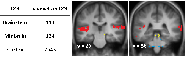
fMRI data were motion corrected, distortion corrected using FSL TOPUP [4,5], RETROICOR physiological noise corrected [6], spatially smoothed (2mm) and analysed in SPM12. Statistical analyses were performed using a GLM of responses to onset and offset, modelled as a stimulus duration=0 at the start/end of broadband noise, and the sustained stimulus, including motion parameters, white matter and CSF as nuisance regressors. A random-effects group analysis was performed to generate SPMs for onsets, offsets and sustained responses using both sampling methods. Region of interest (ROI) masks were formed by thresholding (p<0.001 uncorrected; k=5) SPMs to sustained responses/onsets/offsets for both sampling schemes. Masks were combined (“OR”) to form a single binary mask defining statistically significant sound-related activity which was divided into brainstem, midbrain and cortical ROIs (Figure 1). We evaluate the impact of continuous or sparse sampling on the mean ROI fMRI beta estimates.
Results
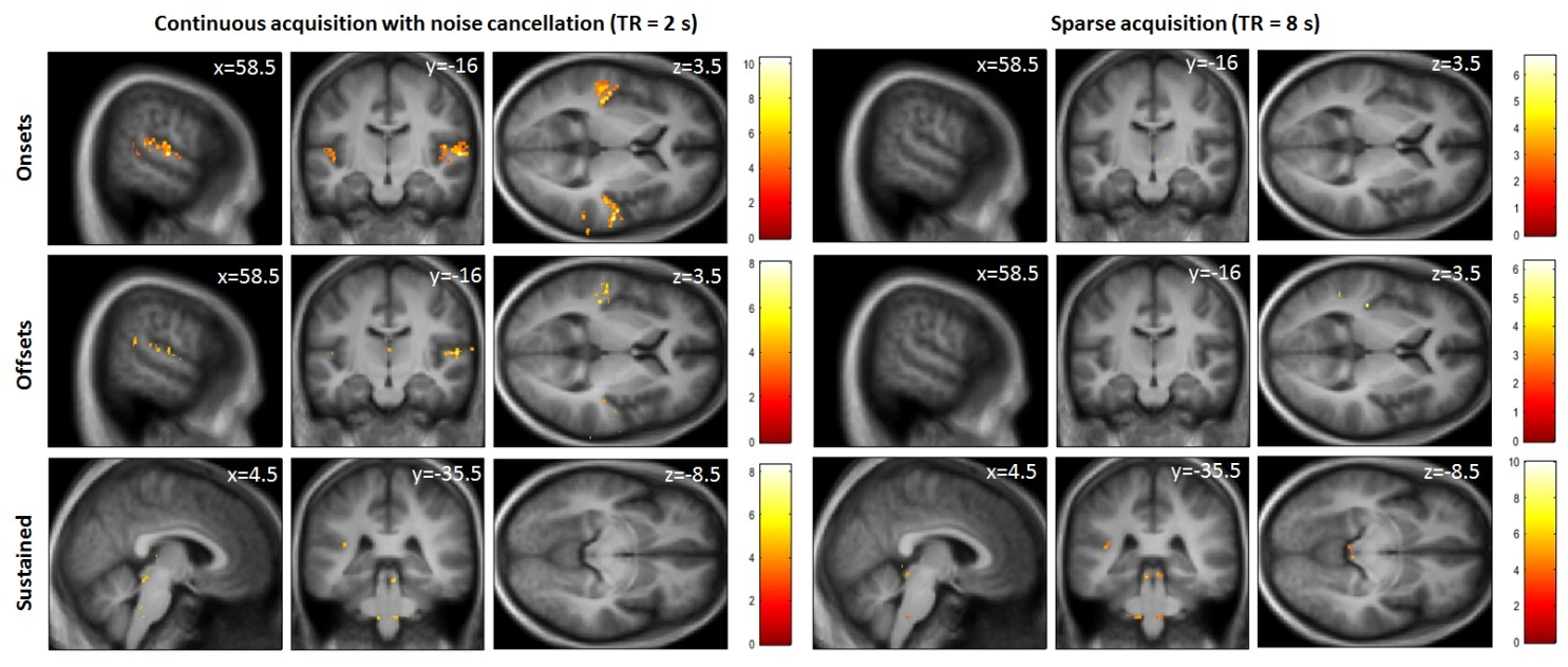
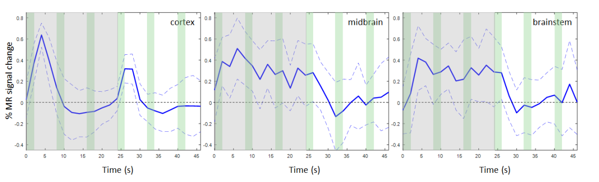
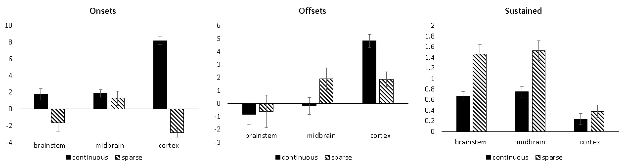
Figure 2 shows group activation maps of the auditory pathway and Figure 3 the timecourse from continuous sampling. Stronger cortical responses to the stimulus onset are seen for continuous sampling. Stronger subcortical responses to the sustained stimulus using sparse sampling. Figure 4 shows the fMRI beta estimates in brainstem, midbrain and cortical ROIs for both sampling schemes. ANOVA statistics showed a significant effect of sampling scheme (F=29.192; d.f.=1,14; p<0.001) and ROI (F=15.755; d.f.=2,28; p<0.001), and significant interactions for ROI*sampling scheme (F=20.348; d.f.=2,28; p<0.001) and ROI*stimulus (F=12.120; d.f.=4,56; p<0.001), demonstrating that the two schemes were selectively preferable for different ROIs of the auditory pathway. As expected, image signal-to-noise ratio (SNR) was higher for sparse than continuous sampling [cortex: 106±12/85±8, brainstem: 111±12/86±8 for sparse/continuous], along with temporal SNR [cortex: 29±1/23±1, brainstem: 14±1/11±1 for sparse/continuous].Discussion
The higher temporal sampling of continuous is superior to sparse sampling for detection of the transient (onset/offset) responses dominant in the auditory cortex. In contrast, sparse sampling was superior to continuous sampling with active noise cancellation in detecting the sustained responses in the subcortical regions. Gains may be explained in terms of reduced interference by scanner acoustic noise and increased SNR from full T1 recovery between acquisitions. It is important to note that a continuous broadband stimulus was selected to stimulate brainstem rather than cortex alone. Future studies will assess the impact of alternative sparse or clustered acquisition timings on functional contrast to maximize both cortical and brainstem responses for sparse sampling.Conclusion
An optimised multiband-sparse fMRI protocol provides significant improvements over multiband-continuous acquisitions to study sustained auditory responses in subcortical regions, even when active noise cancellation is used.Acknowledgements
This work is supported by Medical Research Council (MRC) reference MR/L003589/1 awarded to the University of Manchester.References
1. Harms MP, Melcher JR. Sound repetition rate in the human auditory pathway: representations in the waveshape and amplitude of fMRI activation. Journal of neurophysiology. 2002; 88:1433-1450.
2. Hall DA, Haggard MP, Akeroyd MA, Palmer AR, Summerfield AQ, Elliott MR, Gurney EM, Bowtell RW. "Sparse" temporal sampling in auditory fMRI. Hum Brain Mapp. 1999; 7(3):213-23.
3. Blackman GA, Hall DA. Reducing the effects of background noise during auditory functional magnetic resonance imaging of speech processing: qualitative and quantitative comparisons between two image acquisition schemes and noise cancellation. J Speech Lang Hear Res. 2011; 54(2):693-704.
4. Smith SM., et al., Advances in functional and structural MR image analysis and implementation as FSL. NeuroImage. 2004; 23:S208-S219.
5. Andersson JL., Skare S, and Ashburner J, How to correct susceptibility distortions in spin-echo echo-planar images: application to diffusion tensor imaging. Neuroimage. 2003; 20(2):870-88.
6. Glover GH, Li TQ, Ress D. Image-based method for retrospective correction of physiological motion effects in fMRI: RETROICOR. Mag Reson Med. 2000; 44:162–167.
Figures