3945
T1w MPRAGE transformation based on distortion-matched anatomy for high-resolution fMRIAdnan Shah1, Takashi Ueguchi1, and Guoxiang Liu1
1Laboratory of Brain Function Analysis and Imaging, CiNet, NICT, Suita City, Osaka, Japan
Synopsis
We demonstrate transformation of T1w MPRAGE anatomy into functional space based on DAIREPI T1w-like reconstruction2. This transformation allows comparison of the respective tissue segmentations for highlighting brain regions affected by signal losses and distortions in functional imaging. Effective evaluation of functional imaging at 7T require such an offline quality check procedure to correctly identify the cause of missing hemodynamic activity in high-resolution fMRI at an individual subject-level. This proposed procedure offers a new way to perform analysis of high-resolution fMRI on a high-contrast T1w anatomy and cortical surface in the native EPI space.
Introduction:
High-resolution fMRI1 offers multiple benefits including access to layer-dependent activity, improved accuracy of sampling gray-matter tissues, and reduced contribution of veins and partial volume effects resulting in functional specificity and precise delineation of hemodynamic activity. To perform meaningful interpretations of high-resolution fMRI, it is very important to overcome certain challenges that include distortion-mismatch in functional and structural images and avoid spatial smoothing and resampling of functional volumes. Towards achieving this goal, we developed a technique called DAIREPI2 that allows reconstructing subject-specific T1w-like anatomy and cortical surface from distortion- and resolution-matched IR-EPI for high-resolution fMRI analysis in the native EPI functional space. In this study, we demonstrate the use of DAIREPI2 T1w-like reconstruction to transform the T1w MPRAGE structural scan into functional space. This transformation allows comparison of the respective tissue segmentations for highlighting brain regions affected by signal losses and distortions in functional imaging. Effective evaluation of functional imaging at 7T require such an offline quality check procedure to correctly identify the cause of missing hemodynamic activity in high-resolution fMRI at an individual subject-level. This proposed procedure offers a new way to perform analysis of high-resolution fMRI on a high-contrast T1w anatomy and cortical surface in the native EPI space.Materials & Methods:
A human brain was scanned using 2D multi-shot EPI sequence on a Siemens MAGNETOM 7T scanner with a 32-channel phased array head coil (Nova Medical, MA, USA) to obtain 64 volumes at spatial resolution of 1×1×1 mm3 followed by the same resolution whole-brain two structural scans employing IR-EPI2 sequence using an inversion time of 400 ms. The same multi-shot pulse sequence was used for acquiring both functional and structural scans except differing mainly in TR, FA, and IR. Functional experiment consisted of a standard block design task of checkerboard visual ON/OFF stimulation. In postprocessing, the acquired IR-EPI anatomical data were T1w-like reconstructed and used for fMRI activations overlay2. In this experiment, T1w MPRAGE followed by PDw images from the same subject were acquired with imaging parameters as follows: TR = 2200 ms, TE = 1.65 ms, FA = 5o, Acquisition Matrix = 256 x 256, Resolution = 1 x 1 x 1 mm3, and TI = 1050 ms for T1w image. The T1w-like reconstructed image is added to the denoised IR-EPI image2 to mainly replacing the zero intensity of suppressed white-matter (WM) in IR-EPI image generating a weighted IR-EPI (wIR-EPI) image. Moreover, the acquired T1w MPRAGE images and PDw images were individually denoised3,4,5 prior to division6 and intensity upscaling resulting in a high-contrast-uniform T1w image (uT1w). FSL FLRIT7 was used to co-register and transform the uT1w image into functional space using wIR-EPI as a reference image with a search cost function of correlation ratio and a transform degree of freedom of 12. This transformation results in an anatomical image in functional space referred to as FuT1w.Results and Discussion:
Figure 1 shows fMRI activity overlaid on T1w-like cortical surface (left) and anatomy (middle). Sphere generated from this anatomy is shown to the right. Figure 2 shows the resulting images in ascending order from denoised IR-EPI (I) to T1w-like reconstruction (II), wIR-EPI (III), and the functional-space transformed anatomical image FuT1w (IV). Figure 3 shows the resulting pial-surface (left column) and inner-cortex (right column) generated8 from uT1w and FuT1w. DAIREPI2 offers anatomical imaging in the native EPI space for the analysis and interpretations of high-resolution fMRI data1. High-resolution fMRI analysis requiring the use of high-contrast T1w anatomy for overlay and cortical surface generation can benefit by using wIR-EPI as a reference to transform uT1w anatomy into functional space resulting in FuT1w image. Figure 4 compares the tissue segmentations of IR-EPI and FuT1w. This comparison allow evaluating distortions and signal drop-outs in functional imaging at 7T offline.Acknowledgements
This study was supported in part by Japan Society for the Promotion of Science (JSPS) Grants-in-Aid for Scientific Research “KAKENHI” (Grant Numbers JP26282223 and JP26350471).References
[1] Liu G et al. Block-Interleaved Segmented EPI for voxel-wise high-resolution fMRI studies at 7T. Proc. Intl. Soc. Mag. Reson. Med. 26 (2018) 5450. [2] Shah A et al. Distortion-Matched Anatomical Imaging using Inversion Recovery-Prepared EPI for high-resolution fMRI. OHBM (2018) 1727. [3] Coupe P et al. Medical Image Analysis. 2010;14(4):483-493. [4] Coupe P et al. IEEE Transactions on Medical Imaging. 2008;27(4):425-441. [5] Wiest-Daessle N et al. MICCAI'2008:171-179. [6] Van de Moortele PF et al. Neuroimage. 2009;46(2):432-46. [7] FSL 5.0.7, Analysis Group, FMRIB, Oxford, UK. [8] Shattuck DW and Leahy RM. Medical Image Analysis 2002;8(2):129-142.Figures
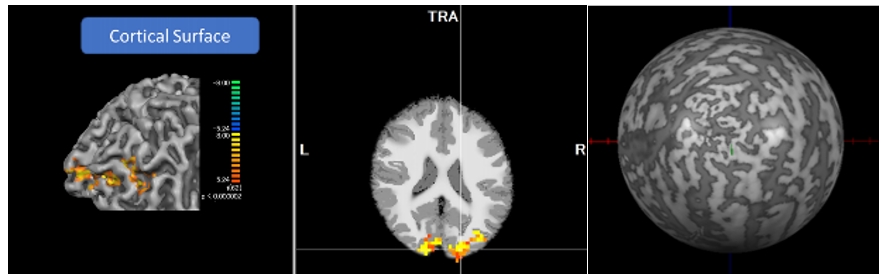
High-resolution fMRI activity overlaid
on T1w-like cortical surface (left) and anatomy (middle). T1w-like sphere
(right).
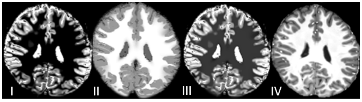
Denoised IR-EPI (I),
T1w-like reconstruction2 (II), wIR-EPI (III), and functional-space transformed uT1w anatomy referred to as FuT1w (IV).
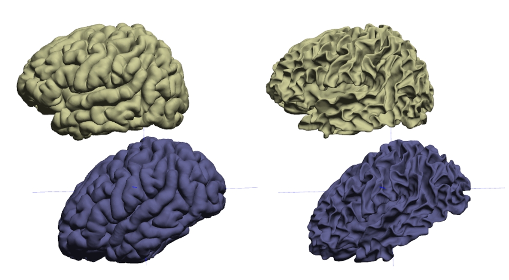
Pial-surface (left) and
the inner cortex (right): generated from uT1w (top) and FuT1w (bottom).

Segmentations overlay FuT1w (red colour) and IR-EPI (yellow colour) for white-matter (1st and 3rd sub-figures) and gray-matter (2nd and 4th sub-figures).