3938
Olfactory functional MRI (fMRI) using T2-prepared BOLD fMRI at ultra-high field (7T)1F.M. Kirby Research Center, Kennedy Krieger Institute, Baltimore, MD, United States, 2Radiology, Johns Hopkins University School of Medicine, Baltimore, MD, United States, 3Neurology, Johns Hopkins University School of Medicine, Baltimore, MD, United States, 4Psychiatry and Behavioral Sciences, Johns Hopkins University School of Medicine, Baltimore, MD, United States, 5Biomedical Engineering, Johns Hopkins University, Baltimore, MD, United States, 6Lou Ruvo Center for Brain Health, Cleveland Clinic, Las Vegas, NV, United States, 7Radiology, Johns Hopkins Hospital, Baltimore, MD, United States
Synopsis
The olfactory cortex is difficult to image with conventional EPI-fMRI due to susceptibility artifacts especially at 7T. T2-prepared (T2prep) fMRI is an alternative method to reduce such artifacts. Here, we show that T2prep-fMRI offers superior temporal signal-to-noise ratio in the olfactory cortex over EPI-fMRI at 7T. The signal characteristics detected using T2prep-fMRI during short (6s) and long (60s) olfactory stimulation were consistent with literature. During the long stimulus, fMRI signals in the olfactory cortex returned to baseline in 5-10s, and activation of secondary olfactory regions (insular, orbitofrontal) were observed, which can be attributed to habituation effects in the olfactory system.
Introduction
The olfactory cortex is difficult to image with conventional EPI-fMRI methods due to significant signal dropout and distortion caused by large susceptibility effects from the nearby temporal bone air especially petrous apex1,2. Such susceptibility artifacts in the olfactory regions (including the entorhinal cortex and parahippocamplal gyrus) have been reported at 3T and are exacerbated at higher field (7T). Recently, a T2-prepared (T2prep) fMRI sequence3-7 was proposed as an alternative fMRI method that can provide images free of signal loss and distortion in regions with large susceptibility artifacts, such as orbitofrontal and olfactory areas close to air cavities, and metallic implants. Here, we applied T2prep-fMRI to detect functional activation in olfactory-eloquent brain regions, and assessed its sensitivity in the olfactory regions. Olfactory functional experiments were performed at 7T with a short and a long stimulation paradigm to evaluate habituation effects in the olfactory system using T2prep fMRI.Methods
All experiments were approved by the local IRB and all participants provided informed written consent. A custom-built multi-channel computer-controlled olfactometer (Whiff LLC, Swarthmore, PA) was used to deliver the odorants in precisely timed pulses. Phenyl ethyl alcohol (PEA) diluted in odorless mineral oil (50% v/v) was embedded in a constantly flowing humidified air stream at body temperature. Each participant was scanned during two different olfactory paradigms: a short stimulation paradigm with 16 blocks of alternating 6s PEA and 30s odorless mineral oil periods (10min); and a long stimulation paradigm with 3 blocks of alternating 60s PEA and 120s mineral oil (10min). fMRI experiments were performed on a 7T Philips scanner using T2prep-fMRI (TR/TE=2.0s/50ms, voxel=1.5mm isotropic, 84 slices, no gap) on 3 healthy subjects. GRE-EPI-fMRI (TR/TE=2.0s/22ms, voxel=1.5mm isotropic, 33 slices, no gap) was also performed on all subjects using only the long paradigm. Data analysis was performed using routines in SPM. A general linear model (GLM) was used for activation detection (adjusted P<0.05).Results
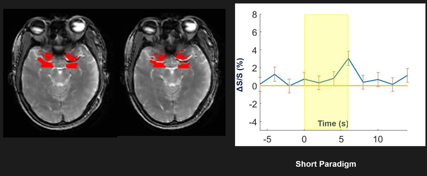
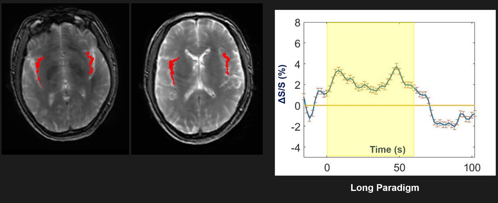
Fig. 1 shows typical images from T2prep- and GRE-EPI-fMRI scans. The red arrows indicate the location of primary olfactory cortex. Temporal signal-to-noise ratios (tSNR) in the olfactory cortex were significantly higher in T2prep scans (105±26) compared to EPI scans (20±14, n=3, P<0.01). Given the higher tSNR in T2prep scans at 7T, the following analysis was focused on T2prep results. Fig. 2 shows a representative activation map in the olfactory cortex detected using T2prep-fMRI during the short olfactory stimulation paradigm. As expected, the time course averaged over activated voxels from all subjects showed a signal increase during the stimulus period. However, little activation was detected in the olfactory cortex during the long paradigm (Fig. 3). The time course averaged over the olfactory cortex showed an initial increase for 5-10s during the stimulus and a quick return to baseline thereafter. Meanwhile, the long paradigm results showed significant activation in brain regions known to be associated with olfaction, including the insula and orbitofrontal cortex. Fig. 4 shows an example of activation in the insula during the long stimulus. Different from the olfactory cortex, the time course averaged over activated voxels in the insula showed elevated signals across the entire period of olfactory stimulation. As activation detection in fMRI is highly dependent on the underlying signal model used in the GLM, we hypothesize that the main reason for the lack of activation in the olfactory cortex during the long paradigm is that the signal model used in the analysis expects a signal increase across the entire period of stimulus, whereas the actual signal increase only occurs in the first 5-10s of the stimulus (green shade in Fig. 5). To show this effect, we repeated the analysis with adjusted time period for signal increase in the GLM, and we found that comparable functional activation in the olfactory cortex can be detected during both the short and long paradigm in the same subjects (Fig. 5).Discussion & Conclusion
T2prep-fMRI showed superior tSNR in the olfactory cortex compared to conventional GRE-EPI-fMRI at 7T. The fMRI signal characteristics detected using T2prep-fMRI during the short and long paradigms were consistent with previous studies in animals8-12 (electrophysiology) and humans13-15. In particular, the quick return of fMRI signals in the olfactory cortex and the activation of secondary olfactory regions during the long paradigm can be attributed to well-known habituation effects in the olfactory cortex13-15. Our data also demonstrate the importance of the underlying signal models in GLM analysis. Further analysis of the data using less constrained approaches16 are underway.Acknowledgements
Funding through the DoD PD160104, NIH R01-NS108452, NIBIB P41 EB015909.References
1. Yang, Q.X., Dardzinski, B.J., Li, S., Eslinger, P.J. & Smith, M.B. Multi-gradient echo with susceptibility inhomogeneity compensation (MGESIC): demonstration of fMRI in the olfactory cortex at 3.0 T. Magn Reson Med 37, 331-335 (1997).
2. Wang, J., et al. Olfactory deficit detected by fMRI in early Alzheimer's disease. Brain Res 1357, 184-194 (2010).
3. Hua, J., Jones, C.K., Qin, Q. & van Zijl, P.C.M. T2- prepared blood-oxygenation-level-dependent (BOLD) fMRI using single-shot 3D fast gradient echo (GRE) sequence with whole brain coverage at 7T. in Proc. 21st Annual Meeting ISMRM 413 (Salt Lake City, USA, 2013).
4. Hua, J., Qin, Q., van Zijl, P.C., Pekar, J.J. & Jones, C.K. Whole-brain three-dimensional T2-weighted BOLD functional magnetic resonance imaging at 7 Tesla. Magn Reson Med 72, 1530-1540 (2014).
5. Hua, J., et al. Robust BOLD activation outside visual and motor cortex during a simple visual and motor task detected by whole-brain T2-prepared spin-echo (SE) BOLD fMRI at 7T. in Proc. 22nd Annual Meeting ISMRM 7382 (Milan, Italy, 2014).
6. Hua, J., et al. Boosting BOLD sensitivity in frontal and temporal regions using T2-prepared BOLD fMRI at 7T. in Annual Meeting OHBM 1859 (Hamburg, Germany, 2014).
7. Hua, J., et al. Language mapping using T2-prepared BOLD functional MRI in the presence of large susceptibility artifacts – initial results in brain tumor and epilepsy patients Tomography 3, 33 (2017). 8. Scholfield, C.N. Electrical properties of neurones in the olfactory cortex slice in vitro. The Journal of physiology 275, 535-546 (1978).
9. Wilson, D.A. Habituation of odor responses in the rat anterior piriform cortex. J Neurophysiol 79, 1425-1440 (1998).
10. Litaudon, P., Datiche, F. & Cattarelli, M. Optical recording of the rat piriform cortex activity. Progress in neurobiology 52, 485-510 (1997).
11. Haberly, L.B. Summed potentials evoked in opossum prepyriform cortex. J Neurophysiol 36, 775-788 (1973).
12. Haberly, L.B. Unitary analysis of opossum prepyriform cortex. J Neurophysiol 36, 762-774 (1973).
13. Georgiopoulos, C., et al. Olfactory fMRI: implications of stimulation length and repetition time. Chemical senses (2018).
14. Sobel, N., et al. Time course of odorant-induced activation in the human primary olfactory cortex. J Neurophysiol 83, 537-551 (2000).
15. Poellinger, A., et al. Activation and habituation in olfaction--an fMRI study. Neuroimage 13, 547-560 (2001).
16. Gonzalez-Castillo, J., et al. Whole-brain, time-locked activation with simple tasks revealed using massive averaging and model-free analysis. Proc Natl Acad Sci U S A 109, 5487-5492 (2012).
Figures