3937
Time variant cardiac noise correction in resting state fMRI without physiologic noise measures1Radiology, Cleveland Clinic, Cleveland, OH, United States
Synopsis
The cardiac and respiratory response functions derived from RETROICOR have substantial variation. We found the cardiac response function is phase, or time shifted across the brain while the respiratory function has a fixed phase, but changes polarity across the brain. Based on this finding, we propose a cardiac and respiratory noise correction method in rsfMRI data without external cardiac and respiratory fluctuation measures, and compare its performance to the RETROICOR model.
Introduction
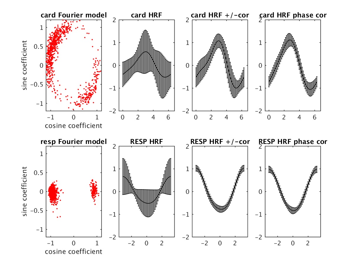
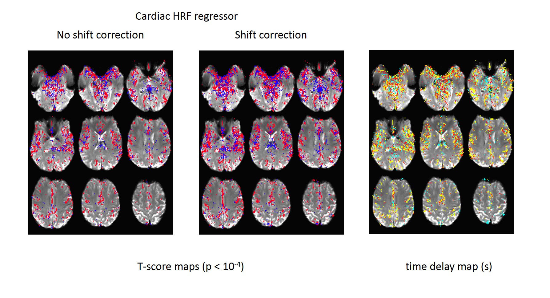
We previously found that the cardiac response function is time-delayed or phase shifted while the respiratory response function has fixed polarity shifts(1), as shown in Fig1. Voxelwise RETROICOR(2) considers time or phase shift of the cardiac and respiratory fluctuations. However any representative cardiac and respiratory signal regressor method including those from PESTICA (3), CORSICA (4), or FIX (5) are limited to detect the time varying cardiac noise. Figure 2 demonstrates the cardiac hemodynamic response function (HRF) calculated from RETROICOR, then cardiac HRF regressor is regressed with and without time shift. As shown, the optimal cardiac HRF regressor varies with a time shift. We propose a cardiac phase shift corrected PESTICA, called as cPESTICA, and validate its performance compared to RETROICOR.Method
Twenty eight healthy controls were scanned at 3T using single band EPI with pulse plethysmograph and respiratory belt recording (TR=2.8s, 128x128 matrix, 31 slices, 132 repetitions). After the first 4 volumes were removed, the second harmonic RETROICOR was applied using modified RetroTS.m (https://afni.nimh.nih.gov/sscc/staff/glend/matlab_compiler). RETROICOR includes the physiologic noise removal process to minimize the error of physiologic regressor model fit. In this study, “RETROICOR model” indicates Fourier series of regressor fitting based on the defined phase. We calculated the voxelwise cardiac and respiratory HRFs from the top 5% of F value voxels in RETROICOR model for the development and validation of the proposed cPESTICA.
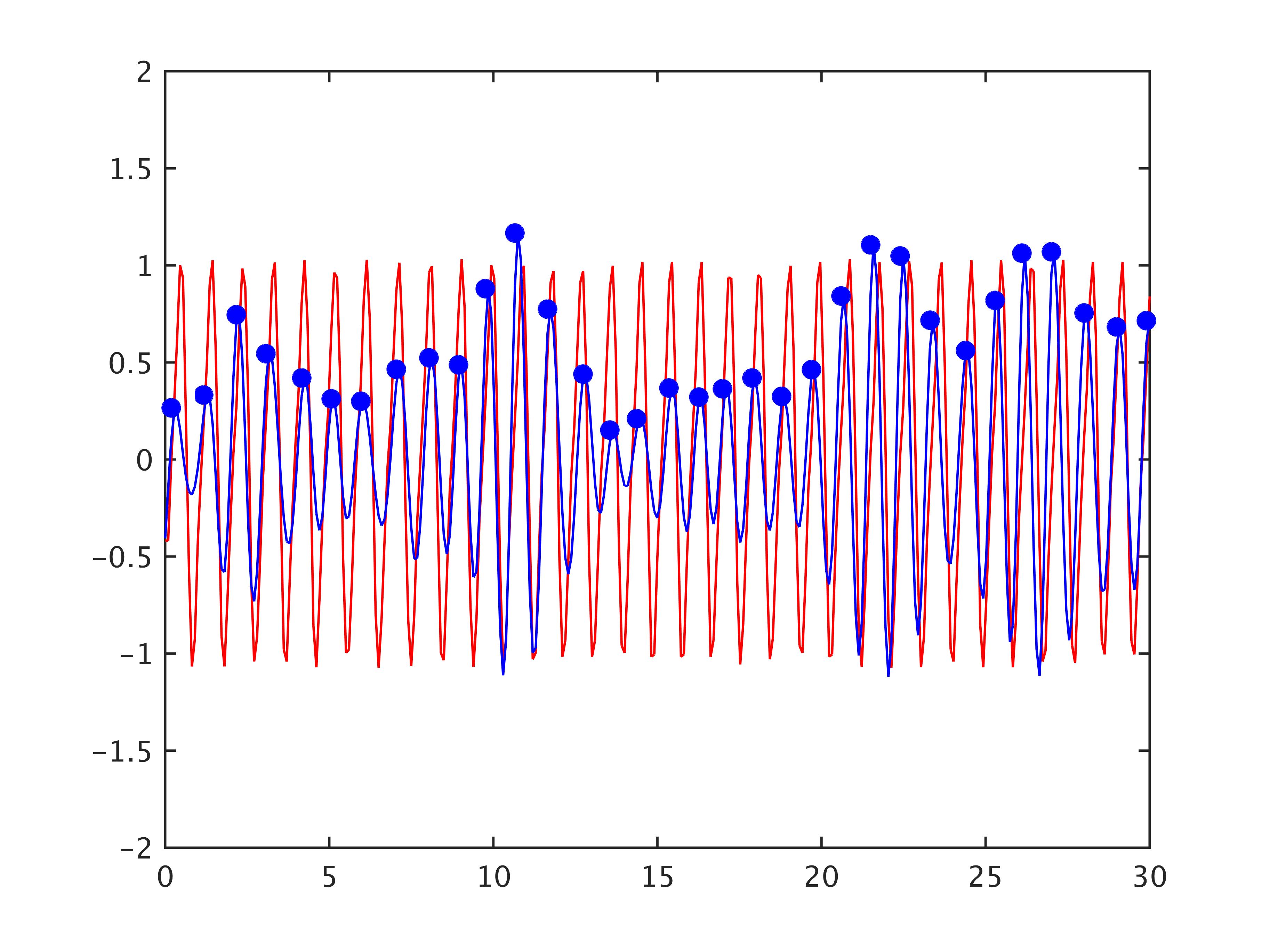
PESTICA generates the physiologic noise estimator using slicewise ICA (3). The first, we define the highest peaks from the estimated cardiac noise estimators from PESTICA, as shown in Fig3, and the linear phase in the time domain from 0 to 2π between the peaks. Note that this phase, defined from PESTICA is not phase from the actual cardiac pulsation measures. The second order Fourier Series were modeled over the defined phase in the same ways that RETROICOR model doesTherefore, cPESTICA has 5 degree of freedom (DOF) while the original PESTICA and RETROICOR have 2 and 8 DOFs.
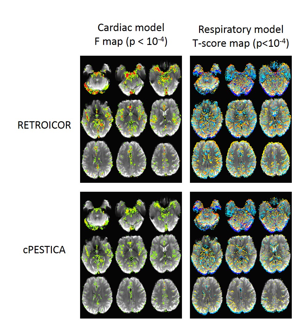
While HRF is NOT used for cPESTICA or RETROICOR correction, HRFs over phase between cPESTICA and RETROICOR are compared for the validation purpose. F value and t-score maps from cPESTICA and RETROICOR are presented.
Result
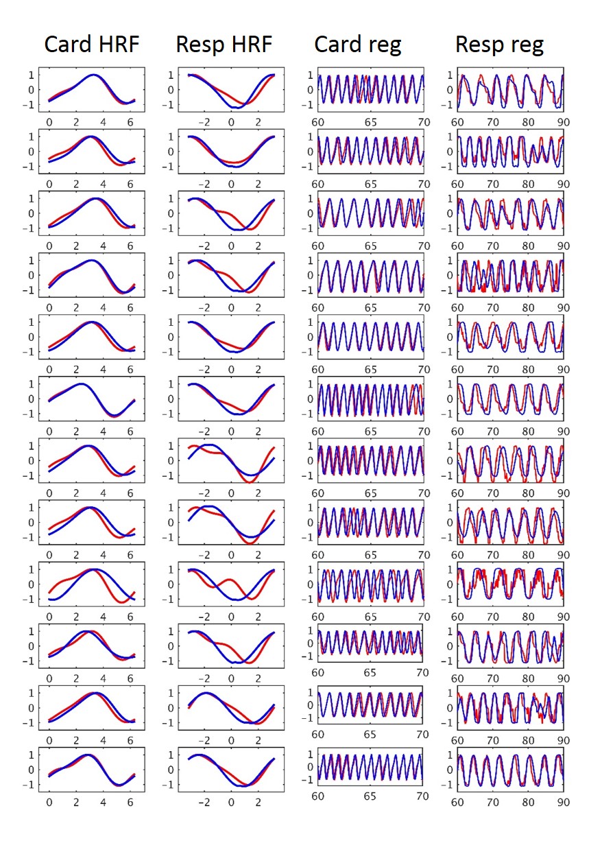
The correlation coefficient (CC) of the cardiac and respiratory HRFs between cPESTICA and RETROICOR are calculated with 0.93±0.08 and 0.90±0.11 across 28 subjects, respectively. CC between the timeseries of cardiac and respiratory HRF regressors are 0.54±0.19 and 0.57±0.17, respectively. Twelve representative HRFs and HRF regressors are presented in Fig4. Fig 5 shows the example of the cardiac and respiratory model fits when using RETROICOR and cPESTICA. The colored areas (p < 10-4) are commonly observed in both RETROICOR and cPESTICA.Discussion
We found that cPESTICA estimates cardiac noise regressors that are equivalent to those produced from the RETROICOR model. Our observed high correlation of the cardiac HRF between cPESTICA and RETROICOR model indicates that cPESTICA can provide reliable physiologic estimators when external physiologic noise measures are not available during fMRI scanning or when the physiologic data is corrupted due to bad contacts or acquisition issues.
This study shows that physiological noise correction with single regressor is only valid for respiratory noise but limited for the time delayed cardiac noise. cPESTICA considers the shift of the cardiac HRF and removes the cardiac noise effectively.
Acknowledgements
Authors gratefully acknowledge technical support by Siemens Medical Solutions.References
1. Shin W, Lowe MJ, editors. A Comprehensive Investigation of Physiologic Noise Modeling in Resting State FMRI; Phase Shifted Cardiac Response Function in EPI. Proceeding of the 26th Meeting of the Society for Magnetic Resonance in Medicine; 2018; Paris, France.
2.Glover GH, Li TQ, Ress D. Image-based method for retrospective correction of physiological motion effects in fMRI: RETROICOR. Magn Reson Med. 2000;44(1):162-7.
3. Beall EB, Lowe MJ. Isolating physiologic noise sources with independently determined spatial measures. Neuroimage. 2007;37(4):1286-300.
4. Perlbarg V, Bellec P, Anton JL, Pelegrini-Issac M, Doyon J, Benali H. CORSICA: correction of structured noise in fMRI by automatic identification of ICA components. Magnetic resonance imaging. 2007;25(1):35-46. PubMed PMID: 17222713.
5. Griffanti L, Salimi-Khorshidi G, Beckmann CF, Auerbach EJ, Douaud G, Sexton CE, Zsoldos E, Ebmeier KP, Filippini N, Mackay CE, Moeller S, Xu J, Yacoub E, Baselli G, Ugurbil K, Miller KL, Smith SM. ICA-based artefact removal and accelerated fMRI acquisition for improved resting state network imaging. Neuroimage. 2014;95:232-47.
Figures