3936
Effect of Coil Compression on tSNR, DVARS and Computation Time on Simultaneous Multi Slice Imaging1Radiology, Medical College of Wisconsin, Milwaukee, WI, United States, 2Center for Imaging Research, Medical College of Wisconsin, Milwaukee, WI, United States, 3Neurosurgery, Medical College of Wisconsin, Milwaukee, WI, United States, 4GE Healthcare, Waukesha, WI, United States
Synopsis
Simultaneous multi-slice (SMS) imaging requires the application of a parallel imaging algorithm for image unaliasing. Including coil compression in SMS image reconstruction offers a benefit of reducing the computational load of the reconstruction algorithm and can better condition the matrix which is inverted in the unaliasing algorithm. The goal of this abstract is to evaluate the optimal level of coil compression to utilize with slice-ARC in Human Connectome Project (HCP) compliant, and other SMS protocols with a Nova Medical 32-channel head coil. It was found that, for all levels of coil compression, application of the compression algorithm yielded a benefit in reconstruction performance. Additionally, it was found that the application of coil compression does not significantly impact the selection of a CAIPI shift factor unless a coil compression of 50% or greater is used.
Introduction
SMS imaging requires the application of a parallel imaging algorithm for image unaliasing. While an increase in the number of distinct coil array elements better conditions the matrix inversion in the unaliasing problem, it also increases the computational burden of the unaliasing algorithm. Field of view shifts from CAIPIRINHA are also applied while acquiring data to further improve the conditioning of the matrix inverted in the unaliasing problem1.Coil compression includes a means to reduce the number of coils to be reconstructed, while yielding synthetic channels with reduced correlation and preserving maximal information.
It was hypothesized that a given level of coil compression will yield image reconstruction quality which is equivalent to reconstruction without compression, and it will require reduced reconstruction time.
Methods
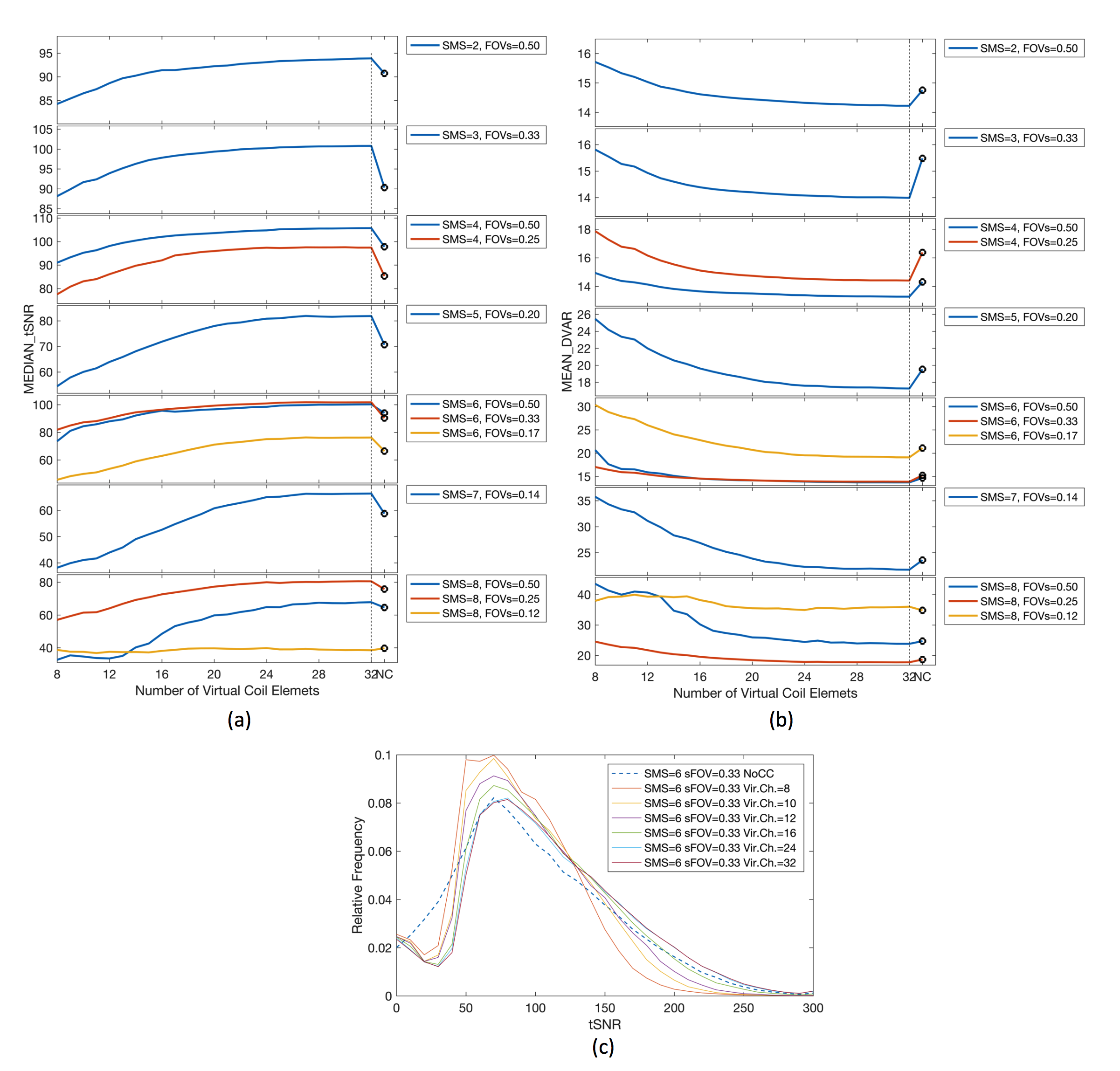
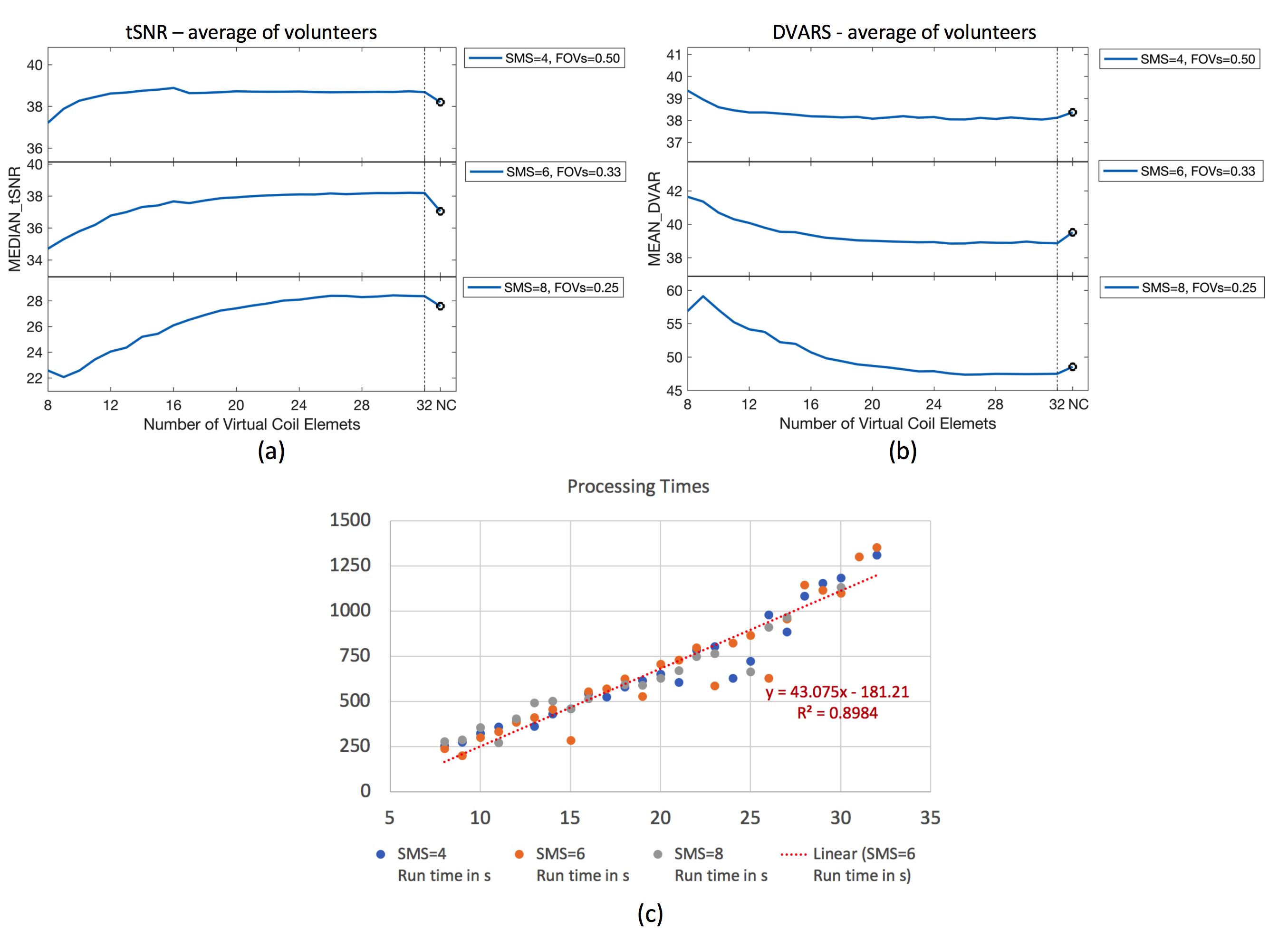
Phantom and human imaging experiments were conducted. In each case, a time series of 114 were acquired with a multi-phase echo planar imaging acquisitions including TE 30ms, TR 1100ms, acquisition duration 2:05, flip angle 50o, and matrix size 104x104. In the phantom acquisition, SMS factors with CAIPI field of view shifts (SMS factor/FOV shift) of 2/0.5, 3/0.33, 4/0.5, 4/0.25, 5/0.2, 6/0.5, 6/0.33, 6/0.17, 7/0.14, 8/0.5, 8/0.25, 8/0.12 were collected. Optimal shifts from the phantom experiment with SMS factors of 4, 6, and 8 were acquired in two consented healthy volunteers (Mean Age:30 years; Mean Weight:155lbs).
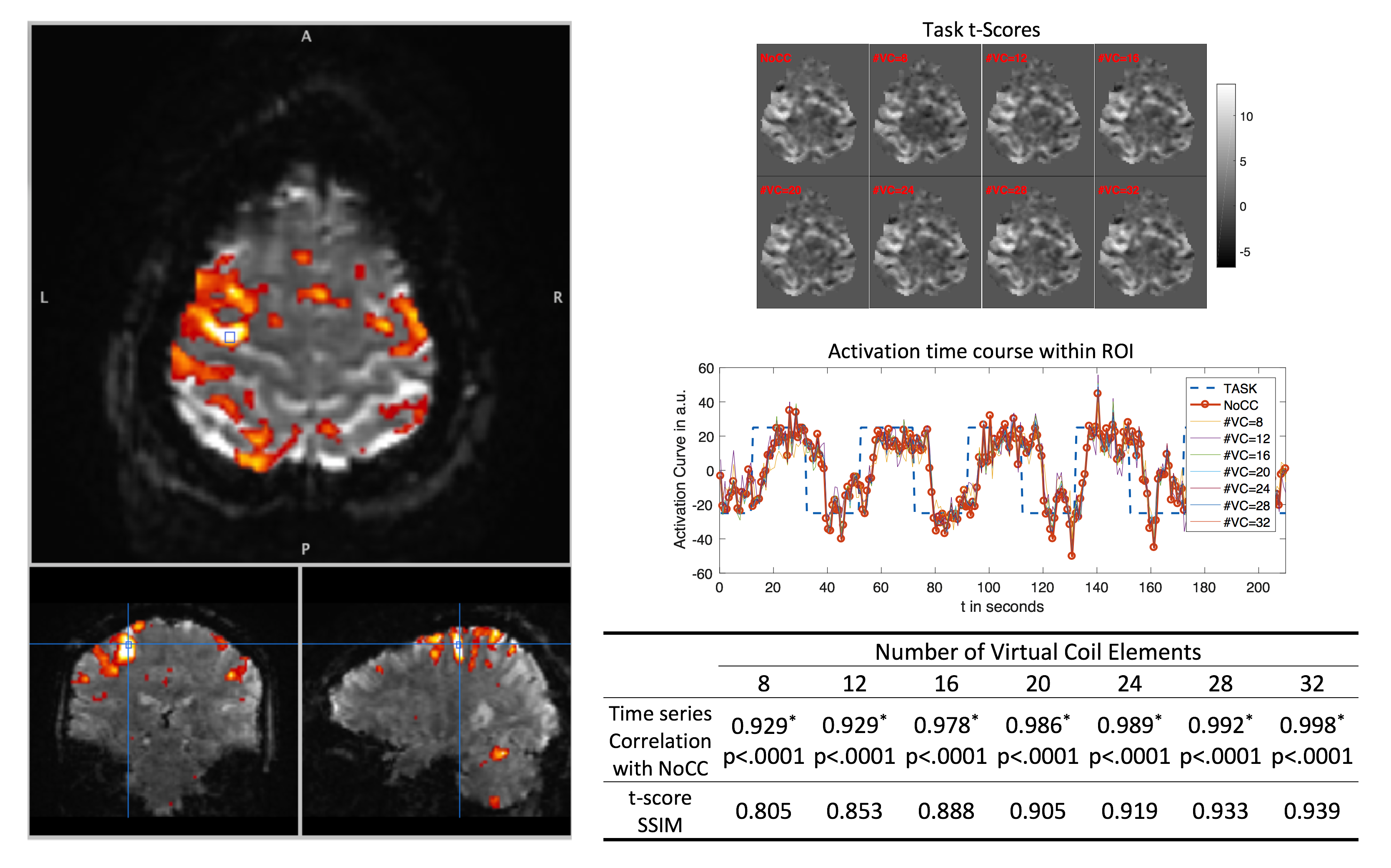
Data were saved for off-line reconstruction wherein an Orchestra C++ algorithm was modified to include coil compression2,3 with different number of virtual coils (range from 8 to 32) before image unaliasing. Reconstruction performance was evaluated via time series temporal signal-to-noise ratio (tSNR—larger values indicate better performance) and temporal derivative of time course of RMS variance over voxels (DVARS—smaller values indicate better performance)3 metrics which are calculated using connectome project’s functional quality pipeline. Additionally a 4 minutes acquisition with right hand finger tapping (20s OFF, 20s ON) were conducted with TR of 0.8s, SMS Factor/FOV shift of 8/0.25 to evaluate the effect of coil compression to task’s t-score maps on the brain and activation curves within 0.042cm3 ROI.
Results
Figure 1 (a) and (b) show the 32-channel Nova head coil receive element coil sensitivities in Axial, Coronal and Sagittal views with a human participant without and with coil compression. This demonstrates that the virtual coil sensitivities are different from the physical coil elements. The total signal with respect to the coil/virtual coil elements are also shown. It is an indicator of the element’s sensitivity to the imaging volume. Figure 1 (c) shows that 22 virtual coils contain over 90% of the image signal. Figure 2 shows the results of the phantom experiment with tSNR increasing with number of virtual coils and DVARS decreasing with number of virtual coils. Of interest, the final point in each plot shows the measurements with no coil compression, yielding compromised metrics compared to reduced numbers of virtual coils. Of further interest, in all cases, the optimal CAIPI shift used for uncompressed coils remained preferred for coil compression factors up to 50%. Figure 3 shows the results for the average of healthy volunteers’ experiments, which are consistent with the results of the phantom experiment. As shown in Figure 3(c), overall processing times are linearly related to the number of coil elements. Processing times for different SMS factors are similar because the FOV coverage and number of slices along with resolution matched. Figure 4 shows activation maps (left), maps of activation t-scores (top right), time series plots in a region of activation (middle right), and a table of correlation coefficients between uncompressed and compressed time series, as well as SSIMs between t-statistic maps without and with compression (bottom right). Activation curves with and without coil compression are extremely similar. All the activation curves are correlated within ROI (Figure 4, Blue Line) with the Pearson correlation coefficient larger than 0.93 (p<0.001) and higher than 0.98 (p<0.001) with coil compression yielding at least 50% of the input channel count. Similarly the task’s t-core maps did not affected by coil compression. If coil compression is 50% or higher, the structural similarity index (SSIM) between the coil compression to without coil compression case is larger than 0.89.Discussion / Conclusions
Coil compression is beneficial in SMS image reconstruction as the reduction in coils reduces computational time, as would be expected and the included image processing yields improved unaliasing performance and image quality metrics. Similarly, it does not have substantial impact of fMRI activation curves and task t-score maps.Acknowledgements
This work supported by GE Healthcare technological development grand and Daniel M. Soref Charitable Foundation.
I would like to thank Dr. Alexander Cohen for his help on fMRI processing and AFNI.
References
1. Setsompop K, Gagoski BA, Polimeni JR, et al., Blipped-controlled aliasing in parallel imaging for simultaneous multislice echo planar imaging with reduced g-factor penalty. Magn Reson Med. 2012; 67:1210-24.
2. Zhang T, Pauly JM, Vasanawala S, Lustig M., Coil compression for accelerated imaging with Cartesian sampling. Magn Reson Med. 2013; 69:571–582.
3. Buehrer M, Pruessmann KP, Boesiger P, Kozerke S, Array compression for MRI with large coil arrays. Magn Reson Med. 2007; 57:1131-1139.
4. Marcus DS, Harms MP, Snyder AZ, Jenkinson M, et. al., Human Connectome Project Informatics: quality control, database services, and data visualization. Neuroimage, 2013; 80:202-219.
Figures
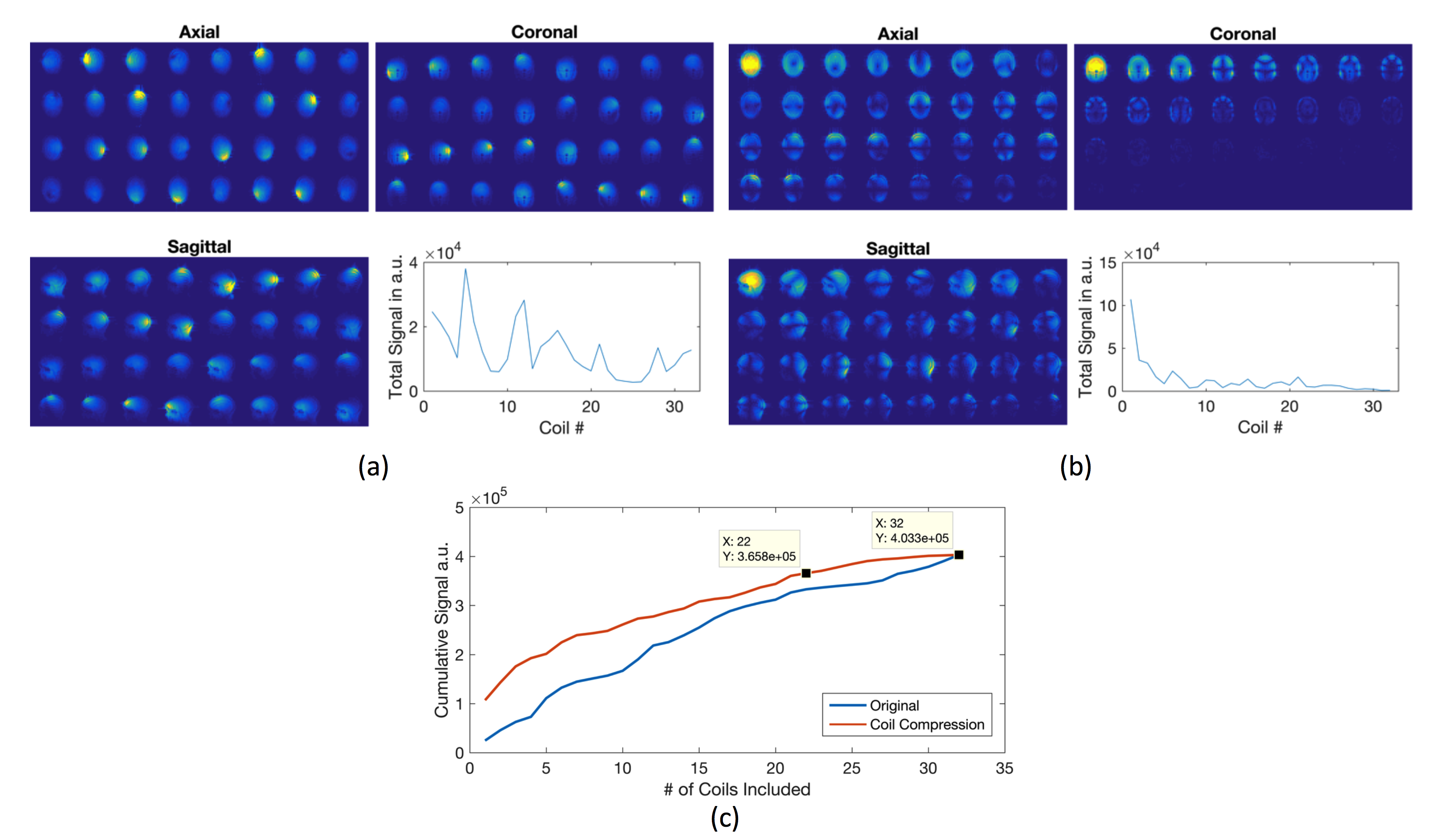
Figure 1. (a) 32 channel head coil receive element sensitivities in orthogonal planes. The plot shows total signal strength, sum of the all the voxels’ absolute values within the FOV, with respect to coil element (arbitrary unit). (b) Virtual elements’ which are sensitized with coil compression algorithm. The plot shows total signal strength (arbitrary unit). Plots in (a) and (b) have the same arbitrary unit. The signal strength has a decreasing trend with respect to virtual element number. (c) Cumulative summation of the coil signal with respect to number of elements. 22 virtual elements are representing almost 90% of the total signal.