3934
Sub-Millimeter Resolution Compressed Sensing GRASE for T2-Weighted Functional MRI at 7 Tesla1University of California, Berkeley, Berkeley, CA, United States, 2Advanced MRI Technologies, Sebastopol, CA, United States
Synopsis
At high magnetic field, 7T and above, T2-weighted fMRI has drawn attention for its high specificity in columnar and layer specific fMRI by minimizing draining vein effects compared to T2*-weighted GE-EPI based fMRI. 3D inner-volume gradient and spin echo (3D-GRASE) has been proposed and used for sub-millimeter resolution. However, reduced field-of-view (FOV) imaging in general has disadvantages compared to GE-EPI: 1) small FOV, 2) T2 induced blurring, both along the partition direction, and 3) low signal-to-noise ratio (SNR) from long TE with EPI readouts. To address both issues, 3D GRASE is extended to volumetric ky-kz random undersampling followed by performing CS for signal recovery (3D CS-GRASE). The ky-kz undersampling extends volume coverage without compromising the blurring while enhancing SNR due to short TE from inplane undersampling.
INTRODUCTION
Functional MRI (fMRI) has been widely used for mapping brain function. At high magnetic field, 7T and above, T2-weighted fMRI has drawn attention for its high specificity in columnar and layer specific fMRI by minimizing draining vein effects compared to T2*-weighted GE-EPI based fMRI1,2. To achieve these benefits, 3D inner-volume gradient and spin echo (3D-GRASE) sequence, which uses slab excitation and refocusing pulses to excite voxels along their intersection using a combination of EPI readouts during multiple spin echoes, has been proposed and used for sub-millimeter resolution3-7. However, reduced field-of-view (FOV) imaging in general has disadvantages compared to GE-EPI: 1) small FOV, 2) T2 induced blurring, both along the partition direction, and 3) low signal-to-noise ratio (SNR) from long TE with EPI readouts. To address both issues, 3D GRASE is extended to volumetric ky-kz random undersampling followed by performing CS for signal recovery (3D CS-GRASE). The ky-kz undersampling extends volume coverage without compromising the blurring while enhancing SNR due to short TE from inplane undersampling.
METHODS
Sequence Design: Accelerated 3D CS-GRASE sequence was designed to permit concurrent acquisition of T2- and T2*-weighted signal components on an EPI readout during fast spin echo acquisitions in the presence of ky-kz random undersampling by placing the central echo of the EPI echo train on the central k-space along the inplane direction. In this manner, upper k-space falls in the first half of EPI echo train and lower k-space in the later half of EPI echo train, respectively. To achieve a high degree of incoherence in time-series data, a ky-kz-t random undersampling was used by distributing the aliasing artifacts along the three dimensions.
Reconstruction: To exploit multi-coil redundancy under the CS framework, distributed CS was used for signal recovery by enforcing joint sparsity instead of individual coil-by-coil sparsity to reconstruct one image time-series that represents the combination from all coils8. For multi-coil combination, coil sensitivities were self-calibrated by averaging undersampled k-space data over time, and a combination of temporal FFT and temporal TV were used as sparsifying transforms.
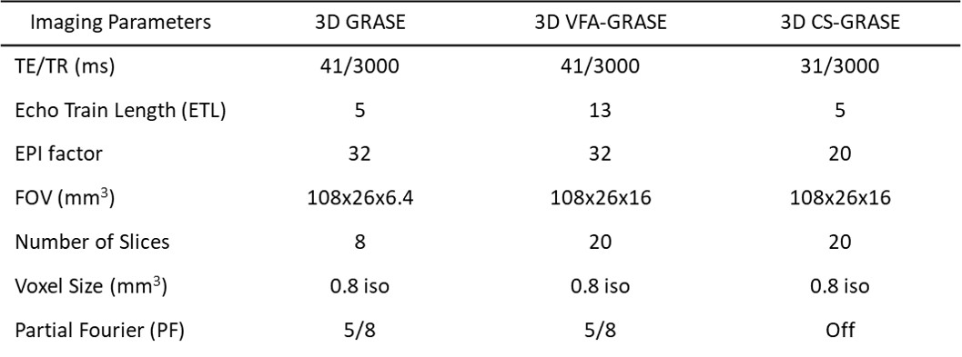
Experiments: All measurements were performed on a 7T MR scanner (Siemens, Germany) with a 32-channel head coil. Informed consent was given by each subject to participate in the protocol according to the institutional review board. Three sets of brain data were acquired using 3D GRASE, 3D VFA-GRASE (variable flip angle)7, and 3D CS-GRASE for evaluation of blurring, temporal SNR (tSNR), and BOLD activation. All imaging parameters were shown in table 1.
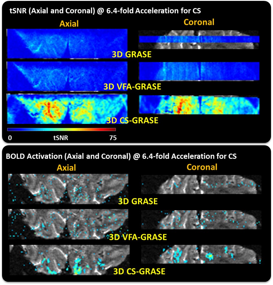
Stimulation Paradigm: BOLD activation was assessed using a vertical meridian localizer stimulus, which alternated between 15s of visual stimulation at the horizontal meridian followed by 15s at the vertical. The cycle was repeated 9 times per scan with on additional half scan at the beginning of the scan to reach steady state.
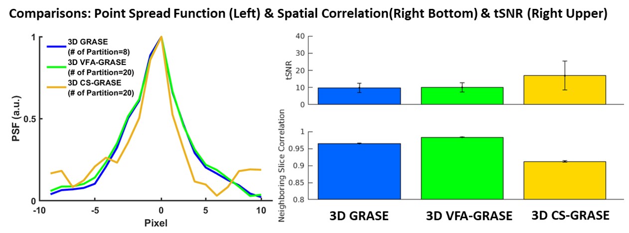
Quality Metrics: Relative image blurring was evaluated using PSF in phantom and spatial correlation9. The spatial correlation between neighboring slices in the temporal mean of each image was compared across the equivalent set of slices, with higher levels of interslice correlation being taken as an indicator of higher levels of blurring. The tSNR was measured by dividing the temporal mean by the temporal standard deviation for each pixel. BOLD activation was measured via a correlation analysis, yielding a coherence value between 0 and 1 indicating the amount of BOLD activity at the frequency of interest.
RESULTS
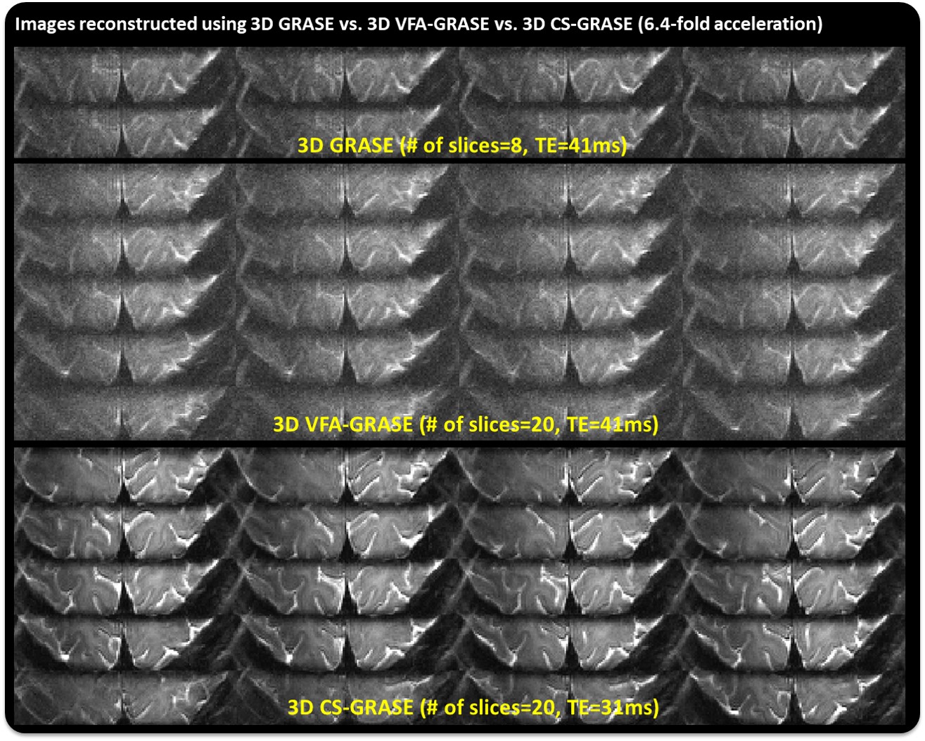
Fig. 1 shows 20 slices acquired in 3D CS-GRASE which is substantially increased from 8 slices in 3D GRASE. The TE is reduced from 41ms to 31ms using CS-GRASE by reducing the EPI factor from 32 to 20, without reducing the in-plane FOV. Fig. 2 demonstrates the 3D CS-GRASE extends volume coverage without compromising the blurring by showing narrower PSF by 1.1 pixel, less interslice correlation by 0.5, and higher tSNR by 10 compared to the competing acquisitions. Fig. 3 shows the corresponding tSNR and its BOLD activation maps. Neither the 3D GRASE nor 3D VFA-GRASE shows significant activations at this resolution with these parameters, whereas the 3D CS-GRASE shows robust activation pattern.
DISCUSSION
Using 3D CS-GRASE to increase the FOV in the partition direction and the shorter TE leads to increased tSNR and BOLD activation. The T2 blurring across slices was slightly mitigated compared to either VFA or standard 3D GRASE and potential optimizations are beyond the scope of this report. These results indicate that CS can be used to extend the FOV of 3D GRASE without inducing additional T2 blurring, and increase the available tSNR relative to 3D GRASE. This presents the opportunity to perform sub-millimeter resolution, T2-weighted fMRI over more extended volumetric coverage than previously available.
Acknowledgements
This work has been supported through NIBIB U01EB025162, NINDS R44NS084788, and NIMH R01MH111444,R24MH106096, R44MH112210.References
1. Yacoub E, Moortele P, Shmuel A, et al. Signal and noise characteristics of Hahn SE andGE BOLD fMRI at 7T in humans. Neuroimage. 2005; 24:738-750.
2. Yacoub E, Shmuel A, Logothetis N, et al. Robust detection of ocular dominance columns in humans using Hahn Spin Echo BOLD functional MRI at 7 Tesla. Neuroimage. 2007;37:1161-1177.
3. Feinberg DA, Harel N, Ramanna S, et al. Sub-millimeter Single-shot 3D GRASE with Inner Volume Selection for T2 weighted fMRI Application at 7 Tesla, ISMRM 2008; p2373.
4. Olman CA, Harel N, Feinberg DA, et al. Layer-specific fMRI reflects different neuronal computations at different depths in human. PLos One. 2012; 7:e32536.
5. Zimmermann J, Goebel R, Maartino F, et al. Mapping the organization of axis of motion selective features in human area MT using high-field fMRI. PLos One. 2011; 6:e28716.
6. Kemper V, Martino F, Vu A, et al. Sub-millimeter T2 weighted fMRI at 7T: comparison of 3D-GRASE and 2D SE-EPI. Front Neurosci. 2015; 9:163.
7. Kemper V, Martino F, Yacoub E, et al. Variable Flip Angle 3D-GRASE for High Resolution fMRI at 7 Tesla. Magn Reson Med. 2016; 76:897-904.
8. Akcakaya M, Hu P, Chuang M, et al. Accelerated Non-Contrast Enhanced PulmonaryVein MRA with Distributed Compressed Sensing. J Magn Reson Imaging. 2011; 33:1248-1255.
9. Vu A, Beckett A, Setsompop K, et al. Evaluation of slice dithered enhanced resolution simultaneous multislice (SLIDER-SMS) for human fMRI, Neuroimage. 2018; 164:164-171.
Figures