3933
Implementing human line-scanning fMRI: Initial results of ultra-high temporal and spatial resolution fMRI1Radiology, University Medical Center Utrecht, Utrecht, Netherlands, 2Spinoza Centre for Neuroimaging Amsterdam, Royal Netherlands Academy of Arts and Sciences (KNAW), Amsterdam, Netherlands, 3Max Planck Institute for Biological Cybernetics, Tuebingen, Germany, 4Graduate Training Centre of Neuroscience, Tuebingen, Germany
Synopsis
Here we present the initial results on implementing human line-scanning fMRI with the purpose of obtaining ultra-high temporal and spatial resolution data of hemodynamic changes in the human brain. This technique could yield novel insights for fundamental neuroscience on laminar information flow, but also for better understanding of microvascular pathophysiological mechanisms in a wide range of brain disorders.
Purpose
Investigate the feasibility of human line-scanning fMRI with the purpose of obtaining ultra-high temporal and spatial resolution data of hemodynamic changes in the human brainBackground
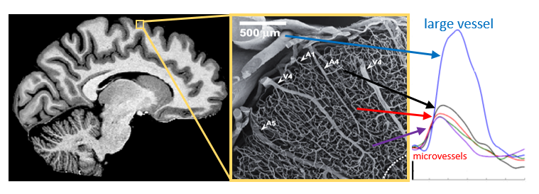
Currently there is an incomplete understanding of how (capillary) blood flow and oxygen distribute across cortical layers to meet the local metabolic demand. Increasing our knowledge on these processes is paramount to advance fundamental neuroscience on laminar information flow, but also for better understanding of microvascular pathophysiological mechanisms in a wide range of brain disorders. There is a rising impetus to identify small vessel damage (microvascular dysfunction) that underly age-related brain disorders1. Noninvasive characterization of microvascular dysfunction will rely heavily on extracting hemodynamic information at very high spatial but also temporal resolution2 (Figure 1). A promising technique that has been pioneered in rodents is the line-scanning fMRI method. The line-scanning approach sacrifices volume coverage and resolution along the cortical surface in order to achieve very high resolution across the cortical depth and time using gradient-echo readouts. The very high temporal resolution, ~50 ms rather than the typical 1-3s in fMRI, will allow filtering out microvessel responses and characterize the distribution and transit of blood flow across the cortical depth. The aim is to extract these responses at a submillimeter (~250um) spatial resolution across cortical depth, but also across hemispheres. Here we report our initial results in implementing the line-scanning method for humans at 7 tesla, comparing resting-state fMRI temporal signal-to-noise ratio measurements and outer volume suppression quality in human primary motor and visual cortex for both a 32 channel head coil and 16 channel surface coil.Methods
Four healthy volunteers were scanned at 7T (Philips) with a 32 channel receive head coil (Nova Medical) and 16 channel high density array surface coil (4). The following line-scanning fMRI acquisition parameters were used: line spatial resolution = 250 µm, TR=50ms, TE=18ms, 2500 timepoints, scan time 4s17min, RF spoiling scheme, flip angle = 16⁰, line length = 180mm, matrix size = 720), line thickness = 2.5 mm, and fat suppression using spectral presaturation with inversion recovery. To obtain a 2.5 mm in-plane line width, two saturation pulses (2.5 mm spatial separation, 2x 6ms pulse duration) were used for outer volume suppression (Figure2). Linescanning was enabled by turning off phase-encoding in the direction perpendicular to the line (no Py). Reconstruction was performed offline using in-house software, multichannel coil data was combined using a sum-of-squares approach before reordering k-space data necessary for line-scanning reconstruction. Temporal signal-to-noise ratios (tSNR) and outer volume suppression quality (ratio signal along the line and signal outside the line) were computed.Results
Suppression of unwanted signals outside the region of the line using outer volume suppression was assessed in a spherical phantom. On average a signal suppression of 96% was achieved (Figure2), in vivo we measured values around 92%. Line-scanning planning, profiles and tSNR in human primary motor (32 channel head coil) and visual cortex (16 channel surface coil) are shown in Figures 3 and 4. For both coils we measured gray matter tSNR ranging between 15-20.Discussion & Conclusion
We presented the initial implementation steps needed to enable gradient-echo line-scanning fMRI for human applications, showing first results in terms acquisition, outer volume suppression, reconstruction, and temporal stability. Adequate outer volume suppression is critical for stable and robust line-scanning fMRI. Our initial findings show signal suppression in phantoms and human subjects ranging between 92-96%. Signal suppression will primarily depend on local B1&B0 which can differ for different regions across the brain, where incomplete suppression can result in bleed-in of unwanted signals reducing tSNR. Next, we will focus on further improving outer volume suppression but also direct spatial excitation for gradient-echo readouts using tailored RF pulses with improved B1&B0-insensitivities. This will likely result in a tradeoff between the time spend on signal suppression and time spend for signal readout (tSNR versus temporal resolution). Another expected tradeoff is the width and thickness of the line (more SNR) versus physiological noise contributions and partial volume effects (reducing tSNR). Other excitation approaches, aimed at spin-echo fMRI readouts, can employ multiple 90⁰ or 90-180⁰ combinations to obtain a pencil-beam excitation, albeit with reduced sensitivity due to stimulated- or spin-echo formation. With these planned improvements and acquisition approaches , tSNR and/or specificity will likely improve enabling task-evoked (functional and hypercapnia) fMRI. Ultimately, the aim is also to acquire line-scanning data at multiple locations along the line and across hemispheres which should be feasible in humans given the highly folded cortical ribbon (see figure 3) in humans compared to rodents.Acknowledgements
This work was supported by a grant of the Royal Netherlands
Academy of Arts and Sciences (KNAW) number: KNAW WF/RB/3781
References
1 Zwanenburg JJM, van Osch MJP. Targeting Cerebral Small Vessel Disease With MRI. Stroke 2017; 48: 3175–3182.
2 Petridou N, Siero JCW. Laminar fMRI: What can the time domain tell us? Neuroimage. Epub ahead of print 20 July 2017. DOI: 10.1016/j.neuroimage.2017.07.040.
3 Yu X, Qian C, Chen D, et al. Deciphering laminar-specific neural inputs with line-scanning fMRI. Nat Methods 2014; 11: 55–8.
4 Petridou N, Italiaander M, van de Bank BL, Siero JCW, Luijten PR, Klomp DW. Pushing the limits of high-resolution functional MRI using a simple high-density multi-element coil design. NMR Biomed. 2013 Jan;26(1):65-73.
Figures