3932
Improving the sensitivity of Spin-Echo fMRI at 3.0 T by highly accelerated acquisitions1Dept. of Radiology, Medical Physics, University Medical Center Freiburg, Freiburg, Germany
Synopsis
Spin-echo fMRI can be highly advantageous compared to gradient-echo fMRI with respect to magnetic field inhomogeneity artefacts, but is seldomly used for BOLD imaging due to its inferior sensitivity. The presented spin-echo implementation of a highly accelerated gradient-echo fMRI pulse sequence therefore aims to improve the sensitivity of spin-echo fMRI while profiting from a reduction of inhomogeneity-induced artefacts. Preliminary measurements using a temporal resolution of 134 ms for whole brain imaging show less signal dropout when compared to the gradient-echo counterpart, as well as an increase in the number of significantly activated voxels compared to the unaccelerated readout.
Introduction
Spin-echo (SE) fMRI can be highly advantageous compared to gradient-echo (GE) fMRI with respect to magnetic field inhomogeneity artefacts such as signal dropouts and geometric distortions [1]. However, at clinical field strengths, the majority of BOLD fMRI experiments are performed using T2* weighted gradient-echo sequences due to their superior sensitivity compared to SE fMRI [2]. In recent years, ultra-fast GE acquisitions using sequences such as simultaneous multi-slice (SMS) or MR-Encephalography (MREG) have been developed that offer the possibility of whole brain imaging in a few hundred ms [3,4]. At such temporal resolutions, benefits include direct filtering of physiological artefacts and improved sensitivities in activation and network connectivity studies. This study therefore aims at the design of a highly accelerated spin-echo-MREG pulse sequence to improve the sensitivity of SE-fMRI while benefiting from a reduction in magnetic field inhomogeneity artefacts.Methods
In order to enable SE-MREG with low flip angle excitation RF pulses (α=25°) [5], two refocusing pulses were implemented into the pulse sequence. This guarantees maximum SNR since a large percentage of the longitudinal magnetization remains along the B0-direction for every excitation. Sample measurements of two healthy volunteers were performed at 3.0 T (Siemens Prisma, Erlangen, Germany) using a 64 RX head coil array. Additionally, GE-MREG, GE-EPI and SE-EPI measurements were performed for comparison of BOLD sensitivity and magnetic field inhomogeneity artefacts. Measurement parameters for SE-/GE-MREG were TR=134ms/TR=100ms, TE=78ms/TE=37ms, matrix size=192×192x150, and spatial resolution=(3×3×3)mm3. A single-shot undersampled non-Cartesian 3D stack of spirals trajectory was used for readout, and data was reconstructed with MATLAB(Natick, Massachusetts, USA) using a conjugate gradient method with Tikhonov regularization [6]. For SE-EPI and GE-EPI, parameters were TR=3980ms/TR=2620ms, TE=75ms/TE=30ms, matrix size 192x192x120/192x192x126, and spatial resolution (3x3x3)mm³. Visual stimulation was performed for one volunteer with alternating 10s flickering checkerboard/20s rest, which was repeated five times. A similar paradigm was used for the other volunteer, who was additionally asked to perform finger tapping at the time of visual stimulation. Following motion correction and smoothing by a 6 mm Gaussian kernel with FSL [7], activation maps were computed using FMRISTAT [8] modelling noise as a 10th-order autoregressive process.Results
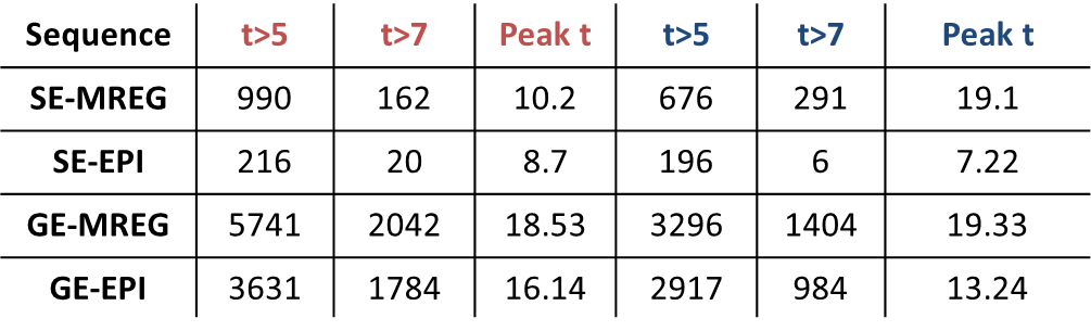
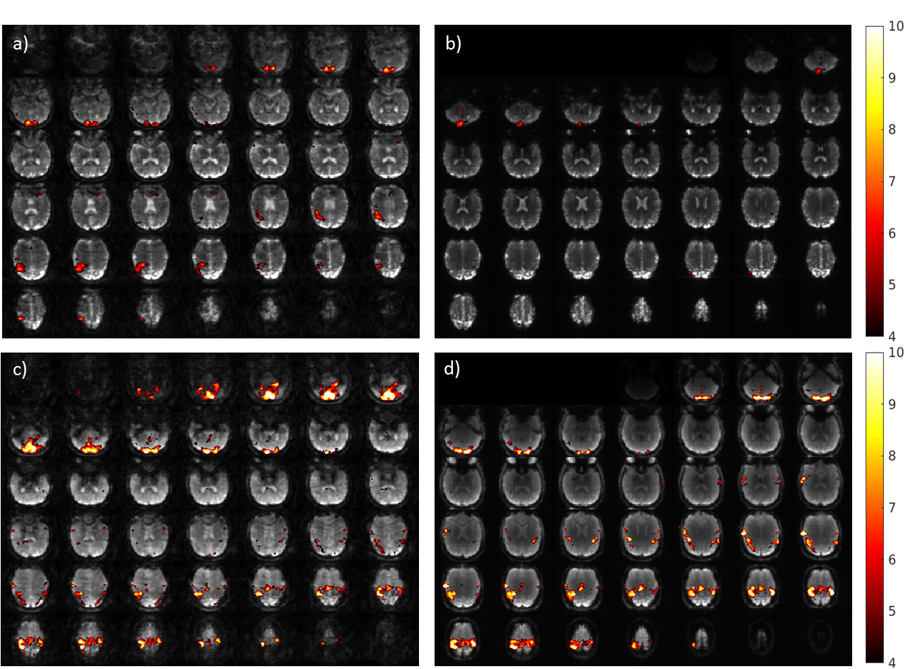
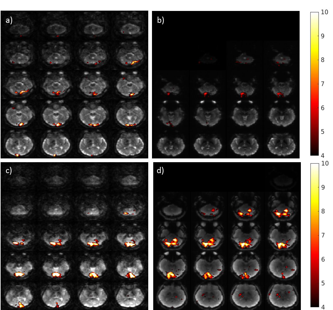
In both fMRI experiments the BOLD sensitivity of SE-MREG was significantly higher than that of SE-EPI, with a 358% and 244% increase in activated voxels above a statistical t-value threshold of t=5 (p<0.05, corrected), respectively (Figure 1). When only considering strong activations at a threshold of t=7, an increase of 4750%/710% was observed. Compared to GE-MREG and GE-EPI, it is more robust to signal dropout in the area of the sinuses (Fig.2 and Fig.3). Blurring artefacts remain due to the long readout of 76ms which enables trajectory distortions before and after the spin echo. The activation maps furthermore revealed a higher sensitivity of GE-MREG compared to GE-EPI in both experiments, with a 43%/14% increase in activated voxels above a threshold of t=7. Although the number of activated voxels of SE-MREG is lower than for both gradient echo pulse sequences, the peak activation value for SE-MREG was higher than that of GE-EPI for the measurement with visual activation only (t=19.31 compared to t=13.24).Discussion/Conclusion
SE-MREG demonstrated a superior sensitivity to SE-EPI in both sample fMRI experiments performed at 3.0 T while additionally benefiting from a high temporal resolution of TR=134ms, suggesting that accelerated acquisitions could constitute a practical approach for SE-fMRI at such magnetic field strengths. Although the overall number of activated voxels was lower than that of both GE-based pulse sequences, likely due to a reduction of intravascular signal in SE fMRI, the local peak activation value was even higher compared to GE-EPI in one of the two measurements. Furthermore, susceptibility-induced signal dropout in the area around the sinuses could be reduced. This offers the opportunity for ultra-fast fMRI in areas strongly affected by magnetic field inhomogeneity artefacts, as for example the inferior temporal lobes [1]. In the future, the present study will focus on implementing an adapted off-resonance correction for SE-MREG. Additionally, sequence parameters such as TR and acquisition time will be adjusted to balance temporal resolution and SNR for optimal BOLD sensitivity.Acknowledgements
This work was supported by grant EXC-1086 „BrainLinks-BrainTools“ from the German Research Foundation (DFG) and grant 13GW0230A from the Federal Ministry of Education and Research of Germany (BMBF).References
1.Halai, A. D., Welbourne, S. R., Embleton, K. & Parkes, L. M. A comparison of dual gradient-echo and spin-echo fMRI of the inferior temporal lobe. Human Brain Mapping 35, 4118–4128 (2014).
2. Norris, D. G. Spin-echo fMRI: The poor relation? NeuroImage 62, 1109–1115 (2012).
3. Feinberg, D. A. & Setsompop, K. Ultra-fast MRI of the human brain with simultaneous multi-slice imaging. Journal of Magnetic Resonance 229, 90–100 (2013).
4. Zahneisen, B. et al. Three-dimensional MR-encephalography: Fast volumetric brain imaging using rosette trajectories. Magnetic Resonance in Medicine 65, 1260–1268 (2011).
5. Tkach, J. A. & Haacke, E. M. A comparison of fast spin echo and gradient field echo sequences. Magnetic Resonance Imaging 6, 373–389 (1988).
6. Assländer, J. et al. Single shot whole brain imaging using spherical stack of spirals trajectories. NeuroImage 73, 59–70 (2013).
7. Jenkinson, M., Bannister, P., Brady, M. & Smith, S. Improved Optimization for the Robust and Accurate Linear Registration and Motion Correction of Brain Images. NeuroImage 17, 825–841 (2002).
8. Worsley, K. J. et al. A General Statistical Analysis for fMRI Data. NeuroImage 15, 1–15 (2002).
Figures