3931
Rotated Stack of Spirals 3D RARE for Single-shot Volumetric ASL Acquisition1Division of MRI research, Radiology, Beth Israel Deaconess Medical Center, Harvard Medical School, Boston, MA, United States, 2Diagnostic Imaging and Radiology, Children’s National Medical Center, Washington, DC, United States, 3Global MR Applications and Workflow, GE Healthcare, Calgary, AB, Canada, 4Global MR Applications and Workflow, GE Healthcare, Boston, MA, United States
Synopsis
Single-shot accelerated volumetric Arterial Spin Labeling is desirable for studies of flow and function modulation but it typically degrades image quality relative to multi-shot interleaved acquisitions. We report a Stack of Spirals RARE acquisition with rotation of spirals between slice encodes and repetitions. This acquisition supports high quality single-shot reconstruction with 3D parallel imaging/compressed sensing while retaining the ability to perform conventional interleaved reconstruction of time averaged perfusion. Application of the sequence to quantification of resting state network fluctuations is demonstrated.
Introduction
Multi-shot interleaved 3D acquisitions with Stack of Spirals (SOS) RARE or GRASE acquisition are widely used for Arterial Spin Labeling (ASL) perfusion imaging because they enable whole-brain coverage, support excellent background suppression, and provide excellent sensitivity. While interleaved acquisitions are suitable for routine time average perfusion measurement, applications and approaches demanding higher temporal resolution are not possible. Stimulated and resting state perfusion fluctuation studies along with dynamic blood flow modulation studies with hypercapnia, blood pressure, or other challenges could benefit from improved temporal resolution through single-shot acquisitions. Acceleration of 3D acquisitions to single-shot would ideally not compromise mean blood flow measurement. Here we report an approximate golden angle spiral rotation strategy compatible with single shot imaging by compressed sensing reconstruction but also standard regridding reconstruction of multi-shot interleaves.Methods
A standard interleaved SOS RARE acquisition sequence was modified in two ways. First the spiral trajectory was modified to variable density for full sampling of the center of k-space in each arm and then 8 fold undersampling of the outer region. Second, the rotation strategy was modified both across shots and slice encodes. Between shots and between increments of slice encoding, the spiral gradients were rotated by an approximate golden angle (Figure 1). This rotation spreads undersampling gaps between the slice and spiral directions to improve parallel imaging while also adding an irregular sampling pattern that may assist with compressed sensing reconstruction. We used an angle of 10π/13, i.e. 138.46°, instead of the golden angle of 137.51°, because the rotation repeats after 13 rotations. The cyclic rotation enables standard slice FFT and 2D regridding reconstruction after 13 interleaves for an excellent quality mean perfusion image that can also be used for coil sensitivity calibration prior to single-shot reconstruction. The repeating rotation pattern also minimizes the memory storage needed for waveforms.
Imaging was performed in 7 healthy volunteers following an IRB approved protocol and written informed consent was obtained prior to imaging. Studies were performed on a GE 3 Tesla MR750 scanner using a 32 channel head array coil from Nova Medical. ASL studies were performed with pseudocontinuous labeling (1.8s labeling, 1.8s postlabeling delay), background suppression, and 32 centric ordered 4mm thick slices. Spiral gradient waveforms were 6.14ms in duration with echo spacings of 12ms. Label and control images were alternated and then rotations of the spiral encode patterns were performed after each pair. 39 rotations were performed corresponding to 3 averages of 13 rotations for a total scan time of 8 minutes.
Images were reconstructed and analyzed off-line. Complex k-space subtraction was performed and fully sampled data were reconstructed by slice direction FFT followed by an in-plane non-uniform FFT (nuFFT). Coil sensitivities were estimated from this fully-sampled perfusion-weighted volume using ESPIRiT(1) (σ=0.01, threshold=0.8) followed by reconstruction of the 39 single-shot volumes using an L1-wavelet Compressed-Sensing reconstruction (λ1=0.005, 100 iterations) with the BART toolbox(2) under MATLAB. Single-shot time series were analyzed for resting state fluctuations(3) using a spatial Independent Component Analysis (ICA) using FASTICA.
Results
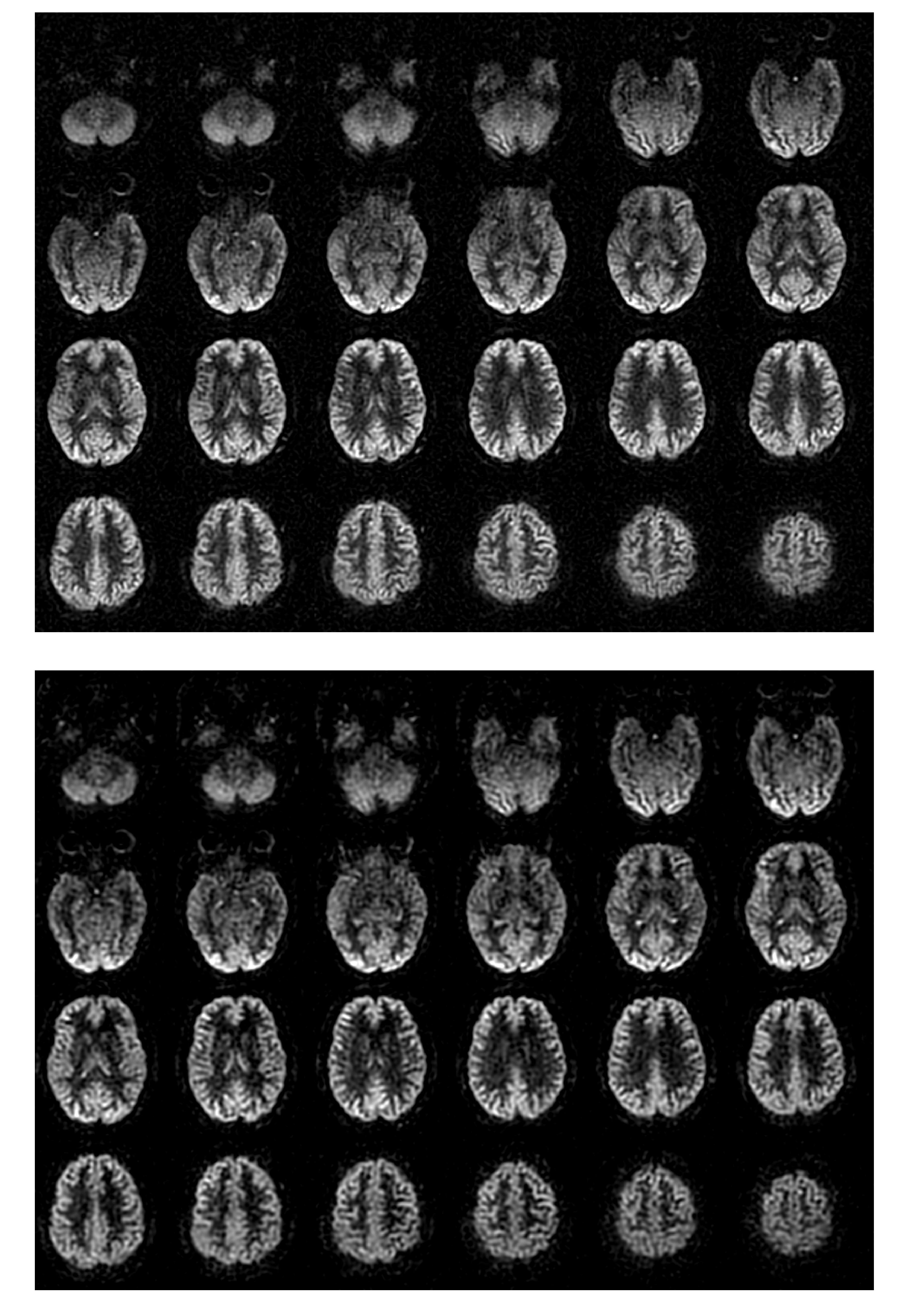
Reconstructed fully sampled images compared favorably with standard interleaved acquisitions acquired with the same ASL labeling parameters, figure 2. 3D CS reconstruction of the 39 individual shots was readily achieved in 1100 seconds on an iMac Pro (6-core Intel Xeon W, 128Gb RAM) with a CPU-based parallelized implementation. Individual single-shot images appeared artifact free but exhibited low SNR consistent with the short acquisition, figure 3. Temporal averages of the single shot images produced images with image quality close to the fully sampled acquisitions, figure 4. ICA analysis of image time series extracted familiar correlated brain networks with good spatial resolution (figure 5) such as default mode, motor and visual networks.Discussion and Conclusions
Building upon prior work using spiral rotations for flexible scan prescription of ASL perfusion(4) and golden angle stack of stars for ASL angiography(5), our approximate golden angle rotation strategy enables uncompromised time average perfusion with the option for accelerated reconstruction up to single-shot with the same data. This should add flexibility for fast ASL, multiple contrast ASL acquisitions such as time-encoded ASL, and studies of perfusion fluctuations and modulation.Acknowledgements
No acknowledgement found.References
(1) Uecker, M. et al. ESPIRiT—an eigenvalue approach to autocalibrating parallel MRI: Where SENSE meets GRAPPA. Magn. Reson. Med. 71, 990–1001 (2014).
(2) Uecker, M. et al. Berkeley Advanced Reconstruction Toolbox. in Proc. Intl. Soc. Mag. Reson. Med 2486 (2015).
(3) Zhao L et al. J Cereb Blood Flow Metab. 2017 doi: 10.1177/0271678X17726625
(4) Li Z et al. Magn Reson Med. 2016 Jan;75(1):266-73
(5) Zhou Z et al. Magn Reson Med. 2017 Dec;78(6):2290-2298
Figures