3930
Quiet, Low-Distortion Whole Brain T2-BOLD fMRI at 7T1MR Applied Sciences Lab West, GE Heathcare, San Francisco, CA, United States, 2Department of Radiology and Biomedical Imaging, University of California, San Francisco, San Francisco, CA, United States, 3MR Applied Sciences Lab Europe, GE Heathcare, Munich, Germany
Synopsis
EPI-based 7T fMRI techniques suffer from spatial distortions in areas of B0 inhomogeneity, signal dropout near air-tissue boundaries, and gradient induced acoustic noise can be as high as 115-120dB. We have developed fast, quiet, low-distortion whole brain 7T fMRI by using the T2-prepared Rotating Ultrafast Imaging Sequence (RUFIS). RUFIS is a ZTE technique with low gradient switching, so it does not suffer from many of the problems of EPI-based methods. It has no spatial distortions and signal dropout, and is 40dB quieter than standard EPI-based technique. Whole-brain 3mm isotropic results for resting state and task-based 7T fMRI experiments are shown.
Introduction
We have developed fast, quiet, and low-distortion whole brain fMRI at 7T using a T2-prepared 3D Rotating Ultrafast Imaging Sequence (RUFIS)1. Because RUFIS is a zero TE (ZTE) imaging technique, it does not suffer from signal dropout near air-tissue boundaries nor from spatial distortions caused by B0 inhomogeneity1,2. Its minimal gradient switching between acquisitions leads to reduced eddy current distortions and low acoustic noise, which are common in EPI-based methods and has been shown to be a confounding factor in fMRI analysis3. This technique has previously been demonstrated at 3T to be 30-40dB quieter than GE and SE-EPI with comparable BOLD sensitivity to SE-EPI2. In this work, we present quiet, whole-brain T2-BOLD 3mm isotropic results for resting state and block design hand-squeezing motor task fMRI experiments at 7T.Methods
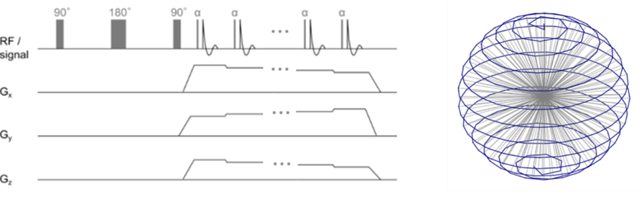
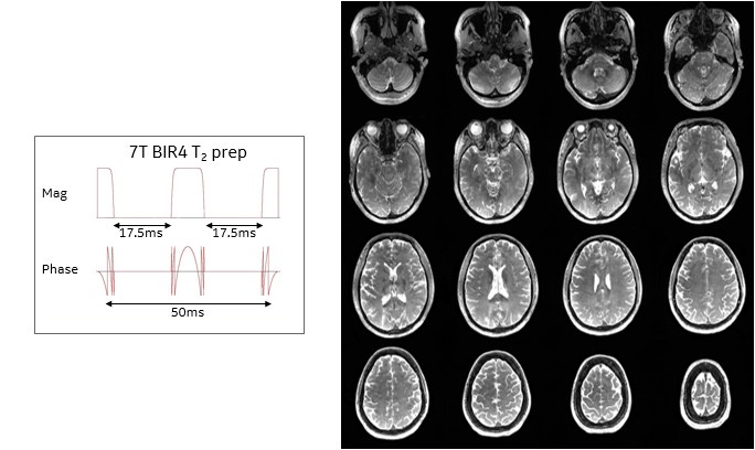
Pule Sequence: The T2-prepared RUFIS fMRI sequence acquires two segments for each volume of k-space. As shown in figure 1, each segment consists of an adiabatic T2-preparation followed immediately by the radial projections in either the top or bottom half of k-space2. The 7T T2-preparation, as illustrated in figure 2, is an adiabatic 0° B1-insensitive-rotation (BIR4) RF pulse4 with a peak B1=9mT, 20ms duration, and 400Hz BW, that was segmented by two 17.5ms intervals to create a T2-prep TEeff=50ms5,6.
Task-based and resting state (rsfMRI) experiments were performed on a GE 7T MR950 scanner (GE Healthcare, Chicago, IL, USA) using a 32Rx/2Tx head coil (Nova Medical, Wilmington, MA, USA). For the task-based experiment, SE-EPI, GE-EPI, and RUFIS fMRI data were collected, and for the rsfMRI experiments GE-EPI and RUFIS fMRI data were collected.
The RUFIS fMRI sequence parameters were: 19.2cm FOV, 64x64x64, 3mm isotropic voxels, FA=2°, BW=±15.6kHz, spokes per segment=512, segments per volume=2, 110 volumes (150 rsfMRI), TR=3s (2.6s rsfMRI), and total scan time=5:30 (7:30 rsfMRI). The data was reconstructed using iterative, non-cartesian SENSE with Total General Variation (TGV) regularization2.
The GE-EPI and SE-EPI fMRI sequence parameters were: 19.2cm FOV, 64x64, 2x1 SENSE, MB1, 64 slices (36 SE-EPI), 3mm slice thickness, 0mm gap, FA=85°, BW=±250kHz, 110 volumes (150 rsfMRI), TE=15ms (50ms SE-EPI), and temporally matched to the RUFIS fMRI.
Experimental Setup: A single, healthy volunteer age 40 years old was scanned for the task-based experiment and seven healthy volunteers ages 22-40 years old were scanned for the rsfMRI experiments. For the task-based experiment, the subject was asked to perform a simple block design task consisting of self-directed left-hand squeezing for 15s followed by 15s of rest for 11 blocks. The subject was visually directed to squeeze their hand and rest using the MR-safe VisuaStim goggles (Resonance Technology Company, Northridge, CA, USA). For the rsfMRI experiment, they were asked to lie awake in the scanner with their eyes closed for the duration of the scan. The experiments were run separately for each of the fMRI sequences described above during the same scan session.
Data analysis: fMRI data were processed using standard preprocessing steps. FSL’s GLM was used to obtain spatial activation maps from the task-based experiment, and FSL's MELODIC was used to extract group-ICA components from the rsfMRI7.
Results
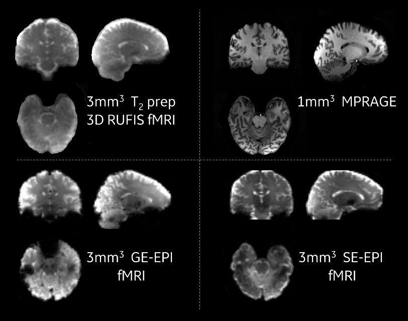
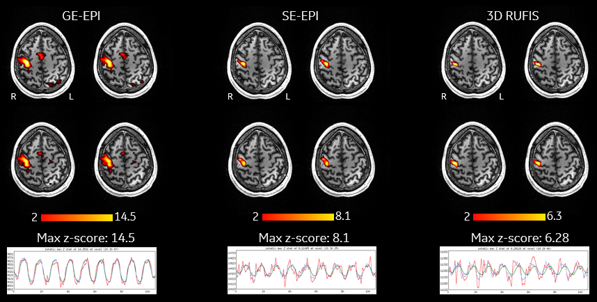
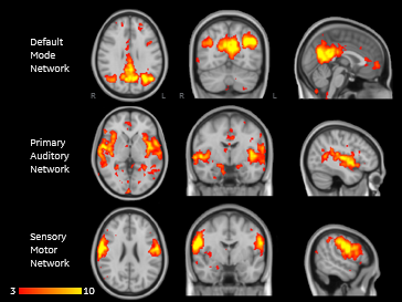
As can be seen in figure 2, the T2 contrast from the 0° BIR4 RF pulse is uniform throughout the brain, including areas of high B0 and B1 inhomogeneity. Figure 3 shows there are no spatial distortions or signal dropout in the T2-prepared RUFIS images, in contrast to the EPI-based techniques. RUFIS shows comparable specificity and T2-BOLD sensitivity to SE-EPI in the contralateral primary motor cortex in figure 4. The RUFIS fMRI sequence had a peak BOLD signal change of 2-3% at 7T, compared to 1-2% at 3T2. The SE-EPI and GE-EPI fMRI sequences had peak BOLD signal changes of 2-3% and 5% at 7T, respectively. The group ICA components from the N=7 RUFIS fMRI resting state scans in figure 5 illustrate that resting state networks can be isolated from this technique. The RUFIS fMRI sequence was barely audible above the ambient noise level in the MRI scan room during each scan session.Conclusion
This preliminary work demonstrates the feasibility of T2-prepared RUFIS fMRI at 7T with nearly twice the sensitivity of this technique at 3T2. It has the potential to deliver many of the benefits of SE-EPI at 7T, such as high CNR and greater specificity than T2*-BOLD8, without many of the drawbacks, such as spatial distortions, signal dropout, and high acoustic noise. This novel, quiet method may enable new auditory experimental paradigms and increase access to subject cohorts with exclusion criteria for EPI-based studies.Acknowledgements
No acknowledgement found.References
[1] Madio DP, Lowe IJ (1995), ‘Ultra-fast imaging using low flip angles and fids’, MRM, vol. 34, 525–529.
[2] Solana AB, Menini A, Sacolick L, Hehn N, Wiesinger F (2016), ‘Quiet and distortion-free, whole-brain BOLD fMRI using T2-prepared RUFIS MR imaging’, MRM, vol 75, 1402–1412.
[3] Moelker A, Pattynama P (2003), ‘Acoustic noise concerns in functional magnetic resonance imaging’, Hum Brain Mapp, vol 20, 123‐141.
[4] Garwood M, Ke Y (1991), ‘Symmetric Pulses to Induce Arbitrary Flip Angles with Compensation for RF Inhomogeneity and Resonance Offsets’, JMR, vol 94, 511–525.
[5] Hua J, Qin Q, van Zijl P, Pekar J, Jones C (2013), ‘Whole-brain three-dimensional T2-weighted BOLD functional magnetic resonance imaging at 7 Tesla’, MRM, vol 72, 1530–1540.
[6] Cao P, Xucheng Z, Shuyu T, Lenyes A, Jakary A, Larson P (2017), ’Shuffled Magnetization-Prepared Multi-Contrast Rapid Gradient Echo Imaging’, MRM, doi:10.1002/mrm.26986.
[7] Jenkinson M, Beckmann CF, Behrens TE, Woolrich MW, Smith S (2012), ‘Fsl’, Neuroimage, vol 6, issue 2, 782-790.
[8] Olman C, Van de Moortele P, Schumacher J, Guy J, Uǧurbil K, Yacoub E (2010), ‘Retinotopic mapping with spin echo BOLD at 7T’, MRM, vol 28, issue 9,1258–1269
Figures