3927
Construction of high precision and resolution render files for SPM and XJview1Center for MRI Research, Academy for Advanced Interdisciplinary Studies, Peking University, Beijing, China, 2Beijing City Key Lab for Medical Physics and Engineering, Institute of Heavy Ion Physics, School of Physics, Peking University, Beijing, China, 3Stanford University, Stanford, CA, United States, 4Department of Linguistics, University of Hong Kong, Hongkong, China, 5McGovern Institute for Brain Research, Peking University, Beijing, China
Synopsis
Render file is one of the typical formats used to display brain surface in brain analysis software such as SPM and XJview. However, current images of render files included in SPM and XJview have issues of low resolution, blurring, and inaccurate spatial localization. Therefore, it is necessary to create a render file with high precision and resolution to better display the activation results generated in SPM. In this study, a set of improved render files were constructed using a modified ICBM_2009c template.
Purpose
To reconstruct a set of high-precision and high-resolution render files to display the activation results generated by SPM more accurately.Introduction
Render file is one of the typical formats used to display brain in brain analysis software such as SPM1 and XJview2. However, current images of render files included in SPM and XJview have issues of low resolution, blurring, and inaccurate spatial localization. Therefore, it is necessary to create a render file with high precision and resolution to better display the activation results generated in SPM. In this study, we first compared the morphological differences between ICBM_2009c(asymmetric)3 and MNI1524 template, and then developed a modified ICBM_2009c template through conscientiously registering the ICBM_2009c_T1 template to the MNI152 space. Based on the modified ICBM_2009c template, a set of high-precision and resolution render files were constructed.Methods
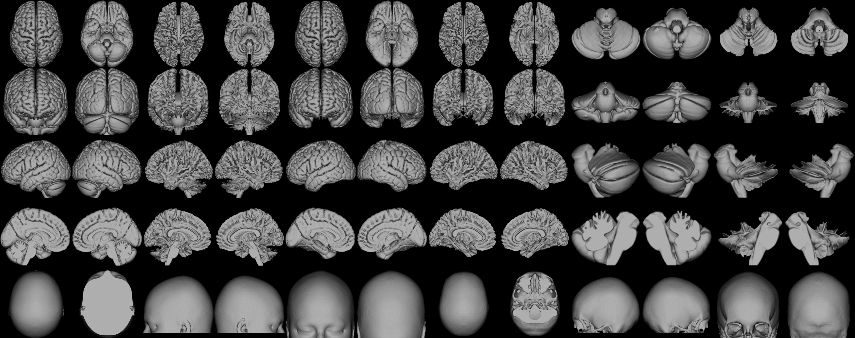
In order to compare the morphological differences between ICBM_2009c and MNI152 template, brain masks were correspondingly produced using the tissue probability maps of these two templates. The subtractive map of two masks can display the morphological difference between two templates. Further, the ICBM_2009c template was normalized to the MNI152 space via the new segment and normalize functions of SPM12, and the image intensity was adjusted for better contrast. Steps to construct the high-precision and -resolution render files are as follows: (1) The grey matter and white matter of the modified ICBM_2009c template were segmented using SPM12. (2) A set of temporary render files were produced by SPM12. (3) The high-resolution brain views of brain and white matter were made by the 3D Slicer software5. (4) The grey matter and white matter views of temporary render files were replaced by the high-resolution brain views that were generated by 3D Slicer. (5) The views of newly temporary render files were recombined into a set of render files with high precision and resolution.Results
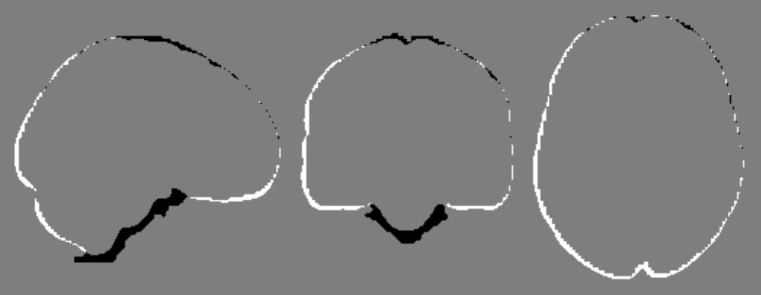
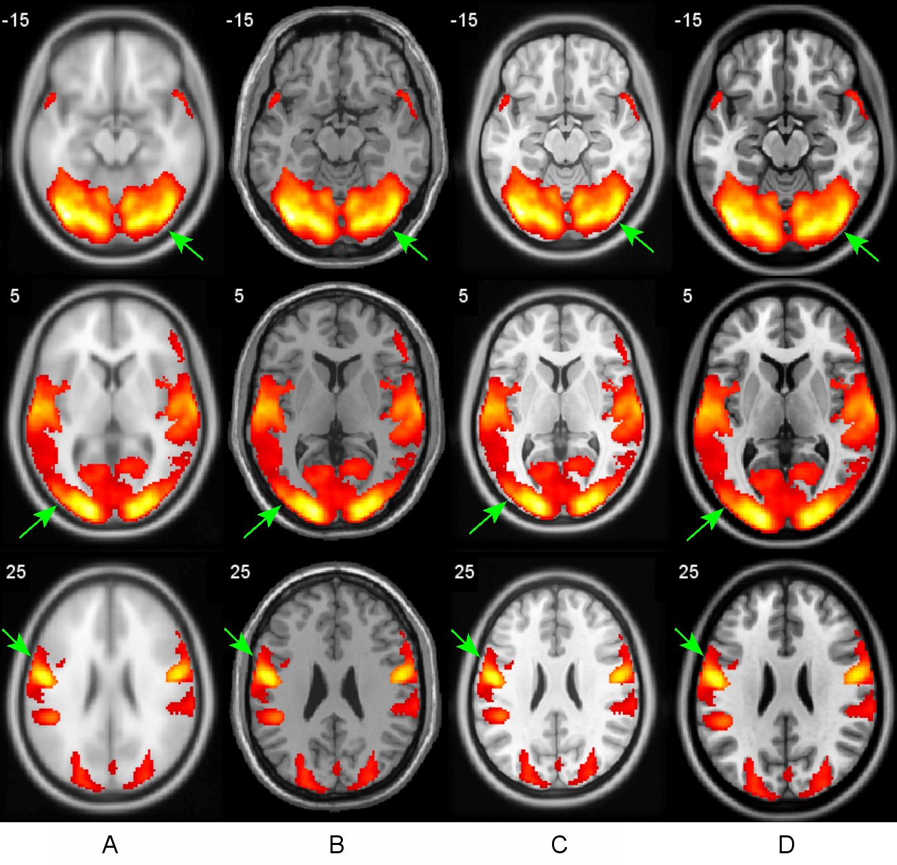
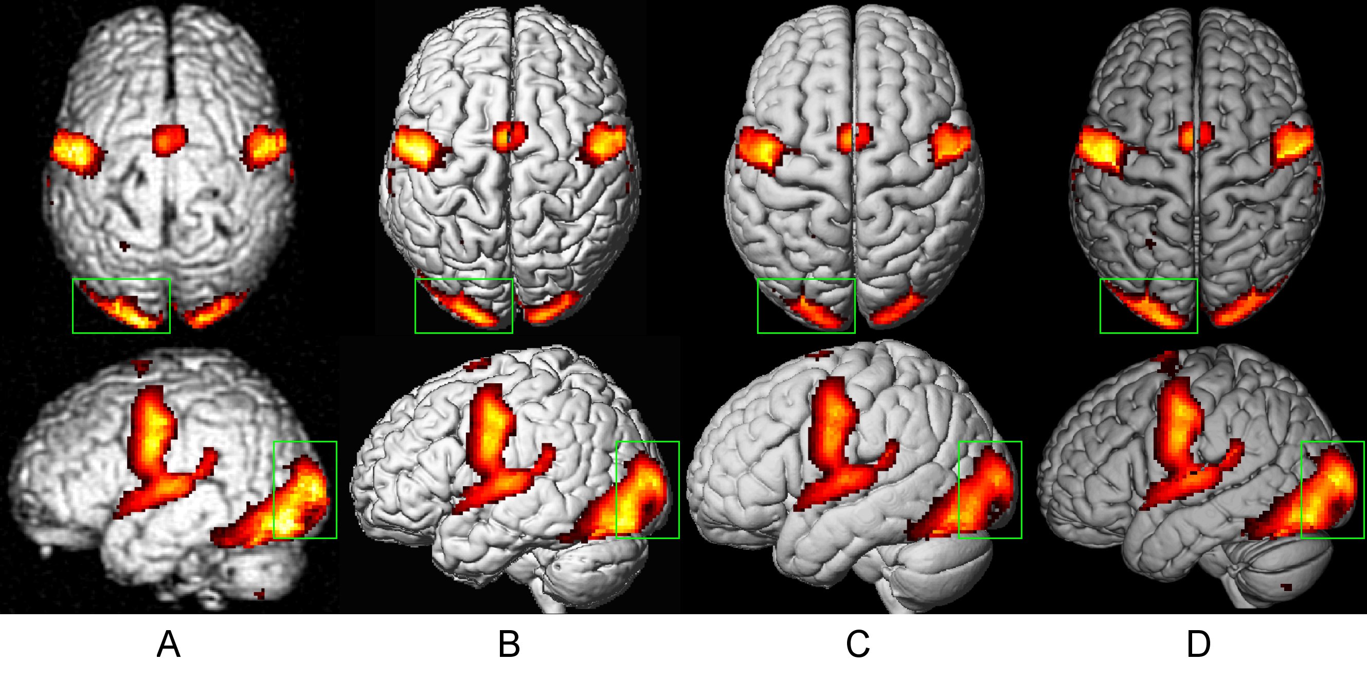
(1) Spatial morphological differences between ICBM_2009c and MNI152 were identified, where the space of ICBM_2009c is slightly larger than that of MNI152 and these two templates own different orientation features (Figure 1). (2) As shown in Figure 2, the new render files have higher resolution and clearer image quality, including the grey matter and white matter surfaces, the cerebrum and cerebellum surfaces, the skull and scalp . (3) To compare the differences of activation display effect between the new template and the old one, activation results generated by SPM12 of a reading task were rendered on different templates. As shown in Figure 3, the activation results show better match with the modified ICBM_2009c template. (4) The activation results of the same task were overlaid on different render files, Figure 4 showed that the images of the newly reconstructed render file are clearer and have higher contrast; also, the activation results are displayed more precisely on the new render file than on others in terms of spatial location.Discussion
Given the spatial differences among different brain templates, mixed use of templates will produce incomparable results due to their uncertain spatial locations, and the positioning of activation results could not be accurately rendered. The render files of SPM have low resolution and poor display effect. The space of XJview (9.6)’s render file is bigger than that of MNI152, and the positioning will be inaccurate as well. Thus, it is necessary to reconstruct a new set of improved render files with high precision and resolution to better display the activation results of SPM.Conclusion
The newly reconstructed render files have the advantages of accurate spatial positioning, high resolution and beautiful display effect. It can be used to display the activation results by SPM and XJview software.Acknowledgements
This study was supported by Beijing Municipal Science & Technology Commission, No. Z171100000117012.References
1. https://www.fil.ion.ucl.ac.uk/spm/
2. http://www.alivelearn.net/xjview/
3. Fonov V, Evans AC, Botteron K, Almli CR, McKinstry RC, Collins DL. Unbiased average age-appropriate atlases for pediatric studies. Neuroimage. 2011-01-01 2011;54(1):313-327.
4. Brett M, Johnsrude IS, Owen AM. The problem of functional localization in the human brain. Nat Rev Neurosci. 2002-03-01 2002;3(3):243-249.
5. https://www.slicer.org/
Figures