3925
Automatic classification of Type 2 diabetes mellitus with and without microangiopathy via feature selection and support vector machine based on resting-state fMRI1Department of Radiology, Shanghai General Hospital, Shanghai Jiaotong University School of Medicine, Shanghai, China, 2Clinical Science, Philips Healthcare, Shanghai, China
Synopsis
Type 2 diabetes (T2DM) mellitus is associated with microvascular complications which can increase risk of cognition impairment and dementia. Recently, machine learning, espicailly support vector machine, were introduced to functional MRI studies in individual classification of diseases. In current study, we used support vector machine to perform individual classification of T2DM with (T2DM-C) and without (T2DM-NC) microangiopathy using ALFF and ReHo features based on rs-fMRI data. The selected features were determined to be key features for classification between groups using recursive feature elimination and may be associated with abnormalities of the spontaneous brain activity in T2DM-C
Introduction
Type 2 diabetes mellitus(T2DM) is a metabolic disease and estimated to affect 450 million adults around the world[1]. T2DM is associated with microvascular complications which can increase risk of cognition impairment and dementia[2]. Previous fMRI study have demonstrated that abnormalities of the spontaneous brain activity were observed in T2DM compared with controls [1]. Recently, machine learning were introduced to functional MRI studies in individual classification and prediction of diseases[3]. In current study, we used support vector machine with linear kernel to automatically classify T2DM with (T2DM-C) and without (T2DM-NC) microangiopathy, and to identifying key features or brain areas associated with microangiopathy.Materials and methods
This study was approved by the Ethics Committee of Shanghai General Hospital, 62 T2DM patients (33 T2DM-C and 29 T2DM-NC) as well as 31 age and sex matched healthy controls (HC) were recuited in this study. Resting-state fMRI data of all the subjects were obtained with a 3T MR scanner (Ingenia, Philips Healthcare, Best, Netherlands). Rs-fMRI data were preprocessed by using the DPARSF toobox (http://www.restfmri.net/forum/DPARSF). The Regional Homogeneity (ReHo) and Amplitude of Low-Frequency Fluctuations (ALFF) were calculated based on preprocessed fMRI data. The mean ReHo and ALFF values of 116 brain regions were extracted based on AAL template and were used as features to classify every two groups (T2DM-NC and HC, T2DM-C and HC, T2DM-NC and T2DM-C). The 10-fold cross validation was applied to generate classification model due to limited sample size. We used recursive feature elimination (RFE) to select features and the survived features were considered as key brain regions in classification process. We used support vector machine (SVM) with linear kernel as the classifier. SVM was an effective and robust classifier to build the model. The linear kernel function were used in this study and it was easier to explain the coefficients of the features for the final model. The model performance was assessed using ROC analysis (receiver operating characteristic). The area under the ROC curve (AUC), accuracy, sensitivity, specificity were also reported in current study. All above processes were implemented with FeAture Explorer (FAE, v0.2.1, https://github.com/salan668/FAE) on Python (3.5.4, https://www.python.org/).Results
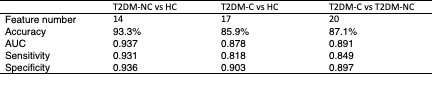
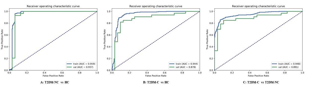
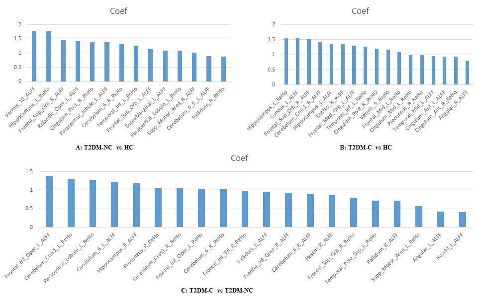
We found that the accuracy and the AUC could achieve 93.3% and 0.937 in T2DM-NC and HC classification using 14 key features, the accuracy and the AUC could achieve 85.9% and 0.878 in T2DM-C and HC classification using 17 key features, the accuracy and the AUC could achieve 87.1% and 0.891 in T2DM-NC and T2DM-C classification using 20 key features, the details presented in Table 1 and Figure 1. The key features in classification of T2DM-NC and T2DM-C exhibited different brain regions compared with classification of T2DM-C vs HC and T2DM-NC vs HC, the details presented in Figure 2.Discussion
In current study, we evaluated the performance of support vector machine to perform individual classification of T2DM-C, T2DM-NC and HC using ALFF and ReHo features based on rs-fMRI data. Our results exhibited that the classification model could achieve higher accuracy (93.33%) when classifying the T2DM-NC and HC groups than other two groups via feature selection. Meanwhile, the classification performance of T2DM-C and T2DM-NC can also achieved good accuracy (87.10%) based on selected features. The survived features were determined to be key features for classification between groups and may be associated with abnormalities of the spontaneous brain activity in T2DM. Especially, the selected features of classifying T2DM-C and T2DM-NC showed different brain regions compared with other two groups, it suggests that the abnormalities of the spontaneous brain activity in T2DM-C exhibited different patterns compared with T2DM-NC and these brain regions may be related with microangiopathy.Acknowledgements
No acknowledgement found.References
[1] . Macpherson H, Formica M, Harris E, et al. Brain functional alterations in Type 2 Diabetes – A systematic review of fMRI studies[J]. Frontiers in Neuroendocrinology, 2017: 34-46.
[2] . Cheng G, Huang C, Deng H, et al. Diabetes as a risk factor for dementia and mild cognitive impairment: a meta-analysis of longitudinal studies.[J]. Internal Medicine Journal, 2012, 42(5): 484-491.
[3] . Billings J M, Eder M, Flood W C, et al. Machine Learning Applications to Resting-State Functional MR Imaging Analysis[J]. Neuroimaging Clinics of North America, 2017, 27(4): 609-620.
Figures