3924
Relative assessment of language lateralisation with fMRI: evaluation of a novel threshold-independent methodIrène Brumer1,2, Enrico De Vita1, Jonathan Ashmore2,3, Jozef Jarosz2, and Marco Borri2
1Department of Biomedical Engineering, School of Biomedical Engineering and Imaging Sciences, King's College London, London, United Kingdom, 2Department of Neuroradiology, King's College Hospital, London, United Kingdom, 3Department of Medical Physics and Bioengineering, NHS Highland, Inverness, United Kingdom
Synopsis
The assessment of language lateralisation with fMRI using the laterality index is limited by the dependence of the results on the chosen activation threshold. To overcome this limitation, different threshold-independent laterality index calculations have been introduced. This work proposes a new method and evaluates how it performs in comparison with three previously reported methods. The methods were evaluated on fifteen healthy subjects who performed picture naming, verb generation, and word fluency tasks. The novel method is simple to implement, fast, robust, reproducible, and compares well with the others in differentiating strong from weak lateralisation on both hemispheric and regional scales.
Introduction
Determining hemispheric or regional dominance in language functions is useful for pre-surgical planning for both brain tumour and epilepsy patients [1,2]. Language lateralisation can be evaluated with functional MRI (fMRI) using the laterality index (LI), which quantifies the dominance of one side of the brain over the other. The conventional LI calculation consists of comparing the number of voxels in the activation map with value above a set activation threshold in left (NL) and right (NR) regions of interest (ROIs): $$$LI=\frac{L-R}{L+R}$$$ . LI values thus range from -1 (right dominant) to +1 (left dominant). This approach is limited by the strong dependence of the LI on the arbitrarily chosen threshold (Figure 1). To overcome this limitation, different threshold-independent LI calculations have been reported [2,3,4,5]. In this work we present a novel threshold-independent method and evaluate its performance against previously proposed methods.Methods
Fifteen right-handed healthy volunteers were scanned following informed consent at 1.5 T (Siemens Aera, standard 20-channel head-only receive coil). The MRI protocol consisted of: 3D T1-weighted MPRAGE anatomical sequence (TE/TR=3.02/2200ms, voxel=(1mm)$$$^3$$$, FA=8°, GRAPPA of 2); fMRI GE-EPI sequences (TE/TR=40/3000ms, voxel=2.5x2.5x3mm$$$^3$$$). fMRI acquisitions consisted of 6 cycles of alternating rest and activation periods of 30 seconds, during which the volunteers performed picture naming, verb generation, and word fluency tasks. Using FSL [6], two different ROIs, created with the Harvard-Oxford Cortical and Subcortical Structural Atlases [7] and Jülich Histological Atlas [8], were considered: 1) the ‘hemisphere ROI’, encompassing the entire cortical hemisphere (excluding the cerebellum) and 2) the ‘language ROI’, defined as the combined Broca's (Brodmann areas 44 and 45) and Wernicke's areas (posterior division of the superior temporal gyrus). Activation t-maps were calculated using SPM12 [9]. The first threshold-independent LI calculation method, labelled ‘curveLI’, calculates the LI as a function of the total number of activated voxels within the ROIs corresponding to different thresholds, and produces a LI curve [1]. The single LI value used for the comparison was calculated at a threshold value corresponding to half the voxels being active (Figure 2). The second method (‘aveLI’) calculates a mean LI by averaging the conventional LI values over the total range of thresholds [4]. The third method (‘histoLI’) integrates the weighted histogram of voxel counts against threshold in right and left ROIs and calculates a global LI [5]. The new method we propose (‘AUCLI’) compares the areas under the curve of the cumulative histogram of voxel counts versus threshold in left and right ROIs, considering all values between 0 and the maximum voxel value (Figure 3). The subjects were ranked according to the LI values obtained for each method. The agreement between pairs of different rankings was quantified by Spearman’s correlation coefficients $$$\rho$$$.Results and Discussion
Figure 4 shows the ranked LI values for all subjects calculated with the four methods for the language ROI. All methods agree on ranking top and bottom subjects and yield similar subject rankings (0.59<$$$\rho$$$<1.00). The ranking comparisons with the histoLI method resulted in the lowest Spearman’s correlation coefficients. While the weighting function of squared t-values used for the histoLI method reduces the influence of low t-value voxels (noise and false positives), it also increases the influence of high t-value voxels, resulting in higher LI values for most subjects or a change of sign (Figure 3, subject 15). Overall, the agreement between methods is higher when the language ROI is considered rather than the hemisphere ROI. However, considering the whole hemisphere might be preferable for patients as accurate localisation of language areas might be difficult due to altered brain pathology and potentially reorganised functionality. The suitability of a LI calculation method for the clinical routine depends on its 1) robustness (independent of any parameter), 2) reproducibility (stability over repeated calculations), and 3) the simplicity of subject comparison [10]. The curveLI, aveLI and AUCLI methods satisfy all three criteria. The histoLI method satisfies criteria 2) only for a determined weighting function. In addition to these criteria, ease of implementation and speed of analysis are important for use in the clinical routine.Conclusion
In this work, we have proposed a new threshold-independent LI calculation method and compared it to three previously reported methods. Our results show the choice of method is not key, as the methods agree in differentiating strong from weak lateralisation on both hemispheric and regional scales, but should be consistent to allow a relative assessment of language lateralisation. This evaluation also suggests that our novel method is well suited for application in the clinical practice as it is simple to implement, fast, robust, reproducible, and allows easy subject comparison.Acknowledgements
This work was carried out at the Department of Neuroradiology at King’s College Hospital NHS Foundation Trust, and supported by the Wellcome EPSRC Centre for Medical Engineering at King’s College London (WT 203148/Z/16/Z) and by the National Institute for Health Research (NIHR) Biomedical Research Centre based at Guy’s and St Thomas’ NHS Foundation Trust and King’s College London. The views expressed are those of the authors and not necessarily those of the NHS, the NIHR or the Department of Health.References
[1] Partovi et al. [2012], ‘Clinical standardized fMRI reveals altered language lateralization in brain tumor patients’, American Journal of Neuroradiology 33(11), 2151-2157. [2] Abbott et al. [2010], ‘fMRI assessment of language lateralization: an objective approach’, Neuroimage 50(4), 1446-1455. [3] Bradshaw et al. [2017], ‘Methodological considerations in assessment of language lateralisation with fMRI: a systematic review’, PeerJ 5, e3557. [4] Matsuo et al. [2012], ‘AveLI: a robust lateralization index in functional magnetic resonance imaging using unbiased threshold-free computation’, Journal of neuroscience methods 205(1), 119-129. [5] Suarez et al. [2008], ‘A surgical planning method for functional MRI assessment of language dominance: influences from threshold, region-of-interest, and stimulus mode’, Brain Imaging and Behavior 2(2), 59-73. [6] Wellcome Centre for Integrative Neuroimaging, FMRIB Analysis Group, University of Oxford, UK [7] Desikan et al. [2006], 'An automated labeling system for subdividing the human cerebral cortex on MRI scans into gyral based regions of interest', Neuroimage, 31(3), 968-980. [8] Amunts at al. [1999], ‘Broca's region revisited: cytoarchitecture and intersubject variability’, Journal of Comparative Neurology, 412(2), 319-341. [9] Wellcome Trust Centre for Neuroimaging, University College London, UK (www.fil.ion.ucl.ac.uk/spm) [10] Nagata et al. [2001], ‘Method for quantitatively evaluating language lateralization of linguistic function using functional MR imaging’, American Journal of Neuroradiology 22(5), 985-99.Figures

Figure 1 : The extent of the activation region depends on the chosen threshold. At low threshold (light red), numerous regions throughtout the brain are considered active but as the threshold increases, only small spots remain (dark blue). The plot shows the dependence of the LI on the threshold.
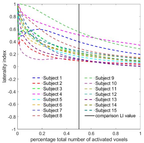
Figure 2: For the curveLI method, the single LI value used for comparison was calculated at a threshold value corresponding to half the voxels being active (mid abscisse).

Figure 3: Number of voxels with value above threshold versus threhsold curves for left and right hemispheres for the verb generation task. For the novel AUCLI method we propose, the areas under these cumulative histograms are used to calculate a laterality index.

Figure 4: Subject rankings obtained with the four methods for the language ROI. The subjects are ordered according to the curveLI mehtod with gradual colour change form blue for low LI values to red for high LI values. The histoLI method presents the worst agreement with the other three methods for all tasks.