3919
An ALE meta-analytic comparison of verbal working memory tasks1Psychology, University of Georgia, Athens, GA, United States
Synopsis
The n-Back and Paced Auditory Serial Addition Test (PASAT) are common verbal working memory tasks (VWM). Activation Likelihood Estimation (ALE) meta-analyses examined n-Back and PASAT literature, revealing regions associated with salience, emotional processing, and VWM. Contrasts revealed differential dorsolateral prefrontal cortex, left posterior parietal cortex, and midline supplementary motor area activation between the tasks. Findings demonstrate the sensitivity of ALE meta-analysis to reveal similarities and differences associated with the cognitive and emotional aspects of VWM tasks. They provide the first glimpse into regions activated by the PASAT using meta-analyses that indicate potential overlapping utility.
Introduction
Verbal working memory (VWM) is a cognitive function responsible for buffering and manipulating recently perceived phonological information that may be transferred to long term memory1. The n-Back and Paced Auditory Serial Addition Test (PASAT) are commonly used VWM tasks that have been used in clinical and experimental settings. Historically, these tasks have been used to assess cognitive impairment in Traumatic Brain Injury, and Multiple Sclerosis2-4 and they have been increasingly employed to induce stress5,6 and examine substance use7-9. However, the literature surrounding the validity of these tasks and their applications differs – the n-Back is better suited for the functional MRI (fMRI) environment with flexibility about difficulty level and stimulus type, while the PASAT possesses norms and is included as part of the Multiple Sclerosis assessment battery3. Some research has indicated that the PASAT is more stressful than the n-Back5,6, yet this has not yet been empirically tested. We employed an activation likelihood estimate (ALE) meta-analysis to determine the extent to which brain regions associated with cognitive and emotional processes are activated by these tasks. We hypothesized that regions associated with VWM would exhibit more activation during the 3-back than the 2-back and PASAT, and that the PASAT would be associated with more activation in emotion-related regions than the n-Back tasks.Methods
ALE10 is a meta-analytic technique that is used to combine and compare results from many studies. This method models the results of prior work by utilizing reported fMRI coordinates of activation to generate a map revealing which regions are most consistently activated across the set of studies. This technique can leverage data that exists in the literature to empirically test hypotheses that were investigated by the individual studies it includes. We compared two difficulty levels of the n-Back task (2-Back and 3-Back) to the PASAT, and to each-other. We used data from published studies that 1) included healthy adults, 2) had more than five participants, 3) employed whole-brain fMRI neuroimaging and 4) reported coordinates for the 2-Back, 3-Back, or PASAT. Cluster-based permutation testing was used for statistical inference, according to the most recent recommendations11-13.Results and Discussion
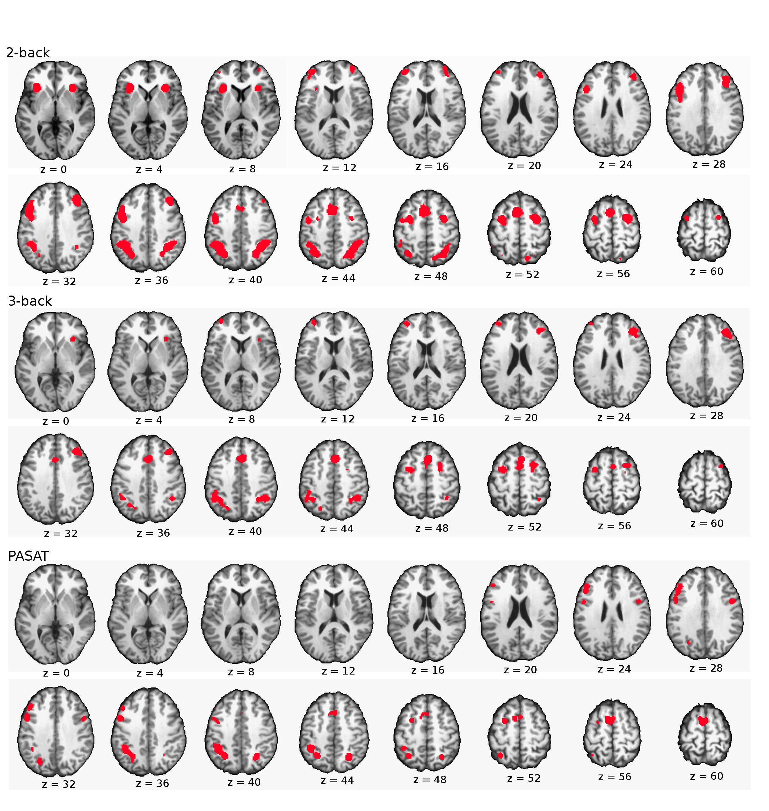
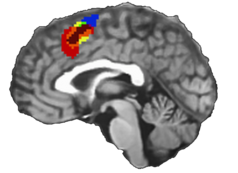
Individual ALE analyses revealed involvement of regions associated with emotional processing and attentional salience (insula, cingulate), in addition to the well-established regions related to VWM (Broca's region, bilateral SMA, premotor, posterior parietal cortices) in all 3 tasks (Figure 1). Overlapping ALE maps revealed an anterior shift in the midline SMA in the 3-Back, while the activity related to the PASAT and 2-back were more posterior (Figure 3). Prior work has suggested that such a gradient may be the result of increasing difficulty14, which may indicate that the PASAT is less cognitively challenging than the 2 and 3-Back.
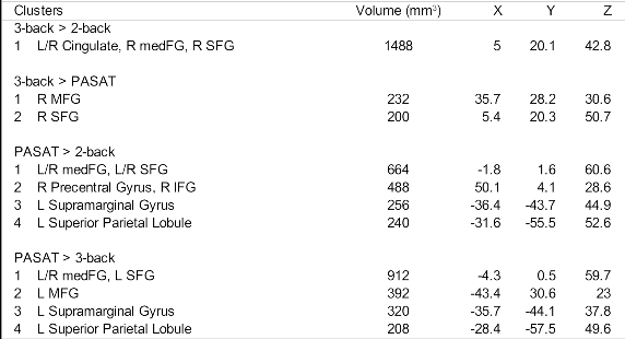
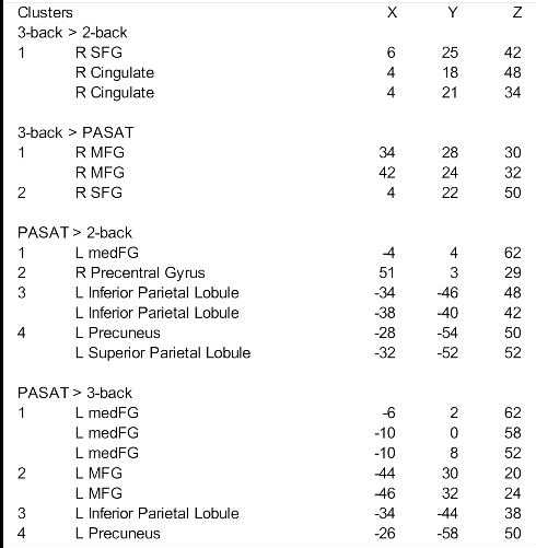
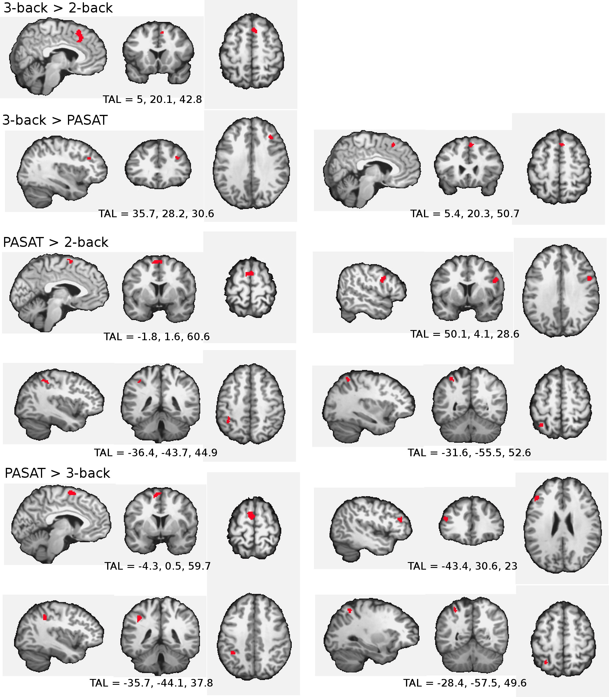
Significant clusters of activation
(Figure 2) were found when contrasting the tasks. Cluster coordinates
and peaks are summarized in Table 1 and Table 2. We observed
lateralization of activation of the DLPFC between the PASAT (left)
and 3-Back (right), which may indicate different cognitive strategies
used to complete these tasks. This lateralized activation in DLPFC
contributes to a growing literature of hemispheric effects in this
region. Furthermore, we saw higher likelihood of activation in the
left posterior parietal cortex during the PASAT, which is involved in
phonological perception, interpretation, and buffering during verbal
working memory. These data suggest greater activation of regions
traditionally associated with the phonological loop during the PASAT,
compared to the 2- and 3-Back tasks.
However, we did not observe differential activation between the
tasks in any emotion-related regions, as we had hypothesized.
Conclusions
These findings are in close agreement with prior meta-analyses that describe regions activated during the n-Back and other working memory tasks15,16. Additionally, these meta-analyses provide the first glimpse into the regions activated by the PASAT, which has not been meta-analytically reviewed prior to this study. These data show specific regions related to cognition and emotion that are activated by these tasks, and highlight the broad similarities between the n-Back and PASAT. Despite significant ALE activation in regions associated with emotion across all tasks, differences between tasks were not observed. This suggests that the tasks are similar in terms of induced stress. Further parametric work examining these tasks is necessary to determine the mechanisms underlying these similarities and differences.Acknowledgements
No acknowledgement found.References
1. Baddeley, A. (2003). Working memory: looking back and looking forward. Nature reviews neuroscience, 4(10), 829-839.
2. Gronwall, D., & Wrightson, P. (1974). Delayed recovery of intellectual function after minor head injury. The Lancet, 304(7881), 605-609.
3. Fischer, J. S., Rudick, R. A., Cutter, G. R., & Reingold, S. C. (1999). The Multiple Sclerosis Functional Composite measure (MSFC): an integrated approach to MS clinical outcome assessment. Multiple Sclerosis, 5, 244-250.
4. McAllister, T. W., Sparling, M. B., Flashman, L. A., Guerin, S. J., Mamourian, A. C., & Saykin, A. J. (2001). Differential working memory load effects after mild traumatic brain injury. Neuroimage, 14(5), 1004-1012.
5. Parmenter, B. A., Shucard, J. L., Benedict, R. H., & Shucard, D. W. (2006). Working memory deficits in multiple sclerosis: Comparison between the n-Back task and the Paced Auditory Serial Addition Test. Journal of the International Neuropsychological Society, 12(05), 677-687.
6. Holdwick Jr, D. J., & Wingenfeld, S. A. (1999). The subjective experience of PASAT testing: Does the PASAT induce negative mood?. Archives of Clinical Neuropsychology, 14(3), 273-284.5.
7. Brown, R. A., Lejuez, C. W., Kahler, C. W., & Strong, D. R. (2002). Distress tolerance and duration of past smoking cessation attempts. Journal of abnormal psychology, 111(1), 180.
8. Daughters, S. B., Lejuez, C. W., Kahler, C. W., Strong, D. R., & Brown, R. A. (2005). Psychological distress tolerance and duration of most recent abstinence attempt among residential treatment-seeking substance abusers. Psychology of Addictive Behaviors, 19, 208 –211.
9. Daughters S. B., Ross T. J., Bell R. P., Yi J. Y., Ryan J., & Stein E. A. (2017). Distress tolerance among substance users is associated with functional connectivity between prefrontal regions during a distress tolerance task. Addiction biology, 22(5), 1378-1390.
10. Turkeltaub, P.E., Eden, G.F., Jones, K.M., Zeffi ro, T.A., 2002. Meta-analysis of the functional neuroanatomy of single-word reading: method and validation. NeuroImage 16, 765– 780.
11. Laird, A.R., Fox, P.M., Price, C.J., Glahn, D.C., Uecker, A.M., Lancaster, J.L., Turkeltaub, P.E., Kochunov, P., Fox, P.T., 2005. ALE meta ‐ analysis: controlling the false discovery rate and performing statistical contrasts. Human. Brain Mapp. 25, 155 – 164.
12. Eickhoff , S.B., Bzdok, D., Laird, A.R., Kurth, F., Fox, P.T., 2012. Activation likelihood estimation meta-analysis revisited. NeuroImage 59, 2349 – 2361.
13. Eickhoff, S. B., Nichols, T. E., Laird, A. R., Hoffstaedter, F., Amunts, K., Fox, P. T., ... & Eickhoff, C. R. (2016). Behavior, sensitivity, and power of activation likelihood estimation characterized by massive empirical simulation. Neuroimage, 137, 70-85.
14. MacKillop, J., Miranda Jr, R., Monti, P. M., Ray, L. A., Murphy, J. G., Rohsenow, D. J., ... & Gwaltney, C. J. (2010). Alcohol demand, delayed reward discounting, and craving in relation to drinking and alcohol use disorders. Journal of abnormal psychology, 119(1), 106.
15. Owen, A. M., McMillan, K. M., Laird, A. R., & Bullmore, E. (2005). N‐back working memory paradigm: A meta‐analysis of normative functional neuroimaging studies. Human brain mapping, 25(1), 46-59.
16. Rottschy, C., Langner, R., Dogan, I., Reetz, K., Laird, A. R., Schulz, J. B., ... & Eickhoff, S. B. (2012). Modelling neural correlates of working memory: a coordinate-based meta-analysis. Neuroimage, 60(1), 830-846.
Figures