3915
Physiological noise removal in fast fMRI without separate physiological signal acquisition1Harvard-MIT Division of Health Sciences and Technology (HST), Cambridge, MA, United States, 2Department of Anesthesia, Critical Care and Pain Medicine, Massachusetts General Hospital, Harvard Medical School, Boston, MA, United States, 3Department of Brain and Cognitive Sciences, Massachusetts Institute of Technology, Cambridge, MA, United States, 4Institute for Medical Engineering and Science, Massachusetts Institute of Technology, Cambridge, MA, United States, 5Department of Biomedical Engineering, Boston University, Boston, MA, United States, 6Department of Radiology, Harvard Medical School, Boston, MA, United States, 7Athinoula A. Martinos Center for Biomedical Imaging, Department of Radiology, Massachusetts General Hospital, Boston, MA, United States
Synopsis
Recent work has shown that commonly used methods to account for physiological noise and serial correlations in conventional fMRI are inadequate for fast (TR<500 ms) fMRI and may lead to incorrect inferences1. We created a model of physiological noise based on harmonic regression with autoregressive noise that utilizes the enhanced sampling of fast fMRI to estimate physiological noise directly from the fMRI data; therefore, it does not require physiological reference signals such as respiration. We found that our model performs as well as gold standard reference-based approaches in removing physiological noise and improves the detection of task-driven fMRI activity.
Introduction
Technological advances in acquisition protocols have enabled an order of magnitude increase in the speed of fMRI measurements2–7. While this new, “fast” fMRI has enormous potential for neuroscientists, the scaling of physiological noise with the improved resolution of fast fMRI limits its applicability8,9. Commonly used pre-whitening and physiological noise regression techniques in conventional fMRI are insufficient to account for serial correlations in fast fMRI, which may lead to errors in interpretation of the fMRI signal1,10. Here we propose a statistical framework that can accurately detect physiological noise sources and separate them from the neurally-driven hemodynamic signal in fast fMRI. Importantly, our approach leverages the enhanced temporal resolution to sample physiological noise directly from the fMRI data, and obviates the need for physiological reference signals (e.g. respiratory belt), which can be technically challenging to collect.
Methods
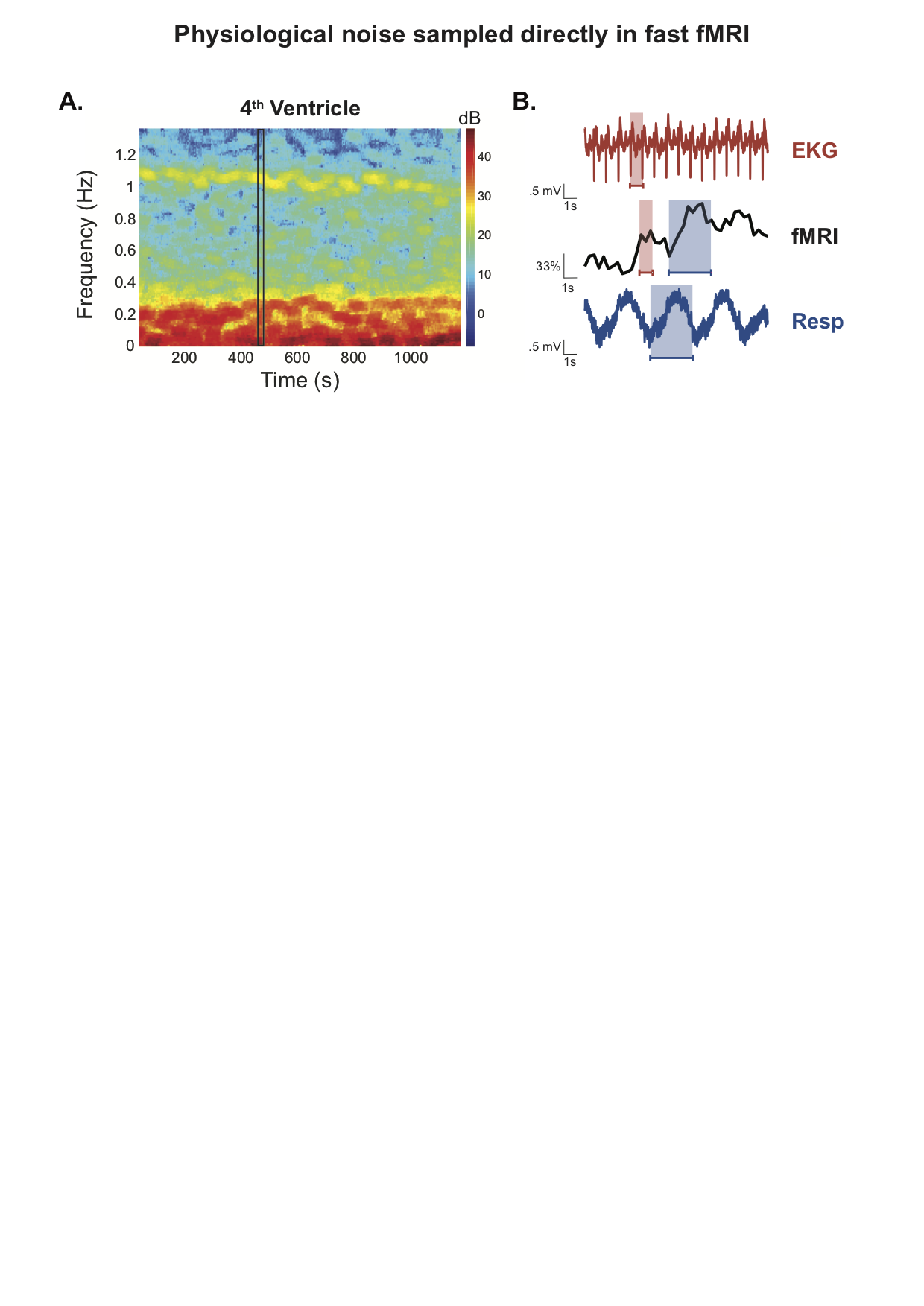
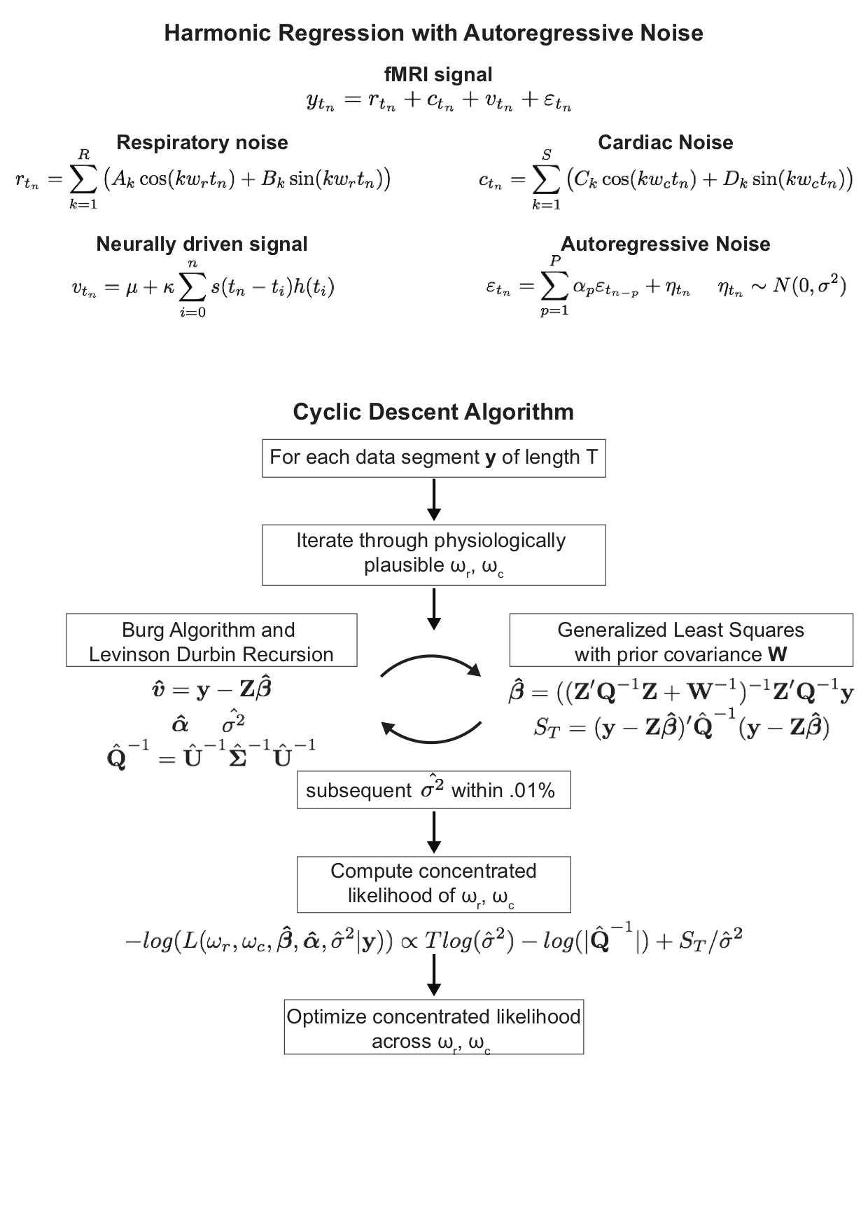
Prior literature and visual inspection of fast fMRI time-series suggest that physiological noise is associated with periodic cardiac and respiratory activity (Figure 1)11. In contrast to conventional fMRI, the temporal resolution of fast fMRI allows direct observation of typical cardiac and respiratory frequencies. Based on these observations and similar models12,13, we designed a model of Harmonic Regression with Autoregressive Noise (HRAN) and an efficient algorithm to compute the maximum likelihood parameter estimates (Figure 2). We first evaluated HRAN performance in a simulated fast fMRI response to a 0.1 Hz stimulus with added cardiac noise (simulated as a 1 Hz sinusoid) and respiratory noise (simulated as a 0.3 Hz sinusoid with one harmonic). Next, we examined HRAN performance in fast resting-state fMRI data collected with physiological reference signals (TR = .367s, 2.5 x 2.5 x 2.5 mm3, 5mm FWHM Gaussian smoothing). Physiological frequency estimates were derived from the mean time series of the 4th ventricle and compared to those obtained from the physiological reference signals. Optimal parameter orders were obtained using the Bayesian Information Criterion (BIC). In the same dataset, the lowest required AR order to remove autocorrelation in each voxel was determined in models with a) no physiological regression, b) physiological regression derived from the fast fMRI data directly using HRAN, and c) physiological regression derived from reference signals using RETROICOR11. Autocorrelations were assessed using the Ljung-Box-Q test. Finally, we evaluated the impact of HRAN on signal detection in a fast fMRI experiment with a 0.1 Hz oscillating visual stimulus for 24s where no reference data was collected (TR = .227s, 2 x 2 x 2 mm3, 5mm FWHM Gaussian smoothing). We derived physiological regressors using HRAN on the left lateral ventricle. We then utilized FSL (fsl.fmrib.ox.ac.uk/fsl/fslwiki/) to obtain activation maps with and without physiological noise removal.Results
In a simulated fast fMRI signal driven by a 0.1 Hz stimulus, HRAN was able to estimate and remove the added physiological noise (Fig 3A,B) and reduce the root mean squared error. In resting-state fast fMRI data, we found that the estimated physiological frequencies derived from the 4th ventricle accurately tracked the average heart rate and respiration rate obtained from the EKG and respiratory belt (Figure 3C,D). HRAN also satisfied goodness of fit criteria with model parameters determined using BIC (Figure 3E). At the single-voxel level, HRAN reduced autocorrelations in the residuals as effectively as RETROICOR (Figure 4A), with greatest impact in gray matter (Figure 4B,C). In a fast fMRI experiment with a 0.1 Hz visual stimulus, HRAN was able to estimate physiological frequencies from the lateral ventricle and improve detection of visually-driven voxels, as compared to standard FSL analysis (Figure 5A-C). For example, in one exemplar voxel, physiological noise modelling with HRAN reduced the residual variance by 56%, enabling detection with a voxel-wise corrected threshold of p < .05 (Figure 5D-F).
Discussion
We found that HRAN is able to accurately estimate physiological frequencies using the fast fMRI data directly and satisfies model goodness of fit criteria. HRAN is as effective as removing autocorrelation as commonly employed techniques such as RETROICOR, while estimating these noise patterns directly from the data itself. These findings suggest that HRAN is able to successfully remove physiological noise from fast fMRI and account for autocorrelation, increasing the accuracy of subsequent statistical analyses.Conclusion
Our model is able to estimate and remove physiological noise from fast fMRI data without the need for physiologic reference signals. Capturing serial correlations using the HRAN framework will not only help to improve interpretations of future fast fMRI experiments, but also help to guide researchers in prospective experimental design. Future work includes implementing time-varying parameter estimates, either by extending our model using state space approaches or incorporating dynamic models such as DRIFTER14.Acknowledgements
This work was funded by NIH grants K99-MH111748, S10-RR023401, and S10-RR023403. Uday Agrawal is a Howard Hughes Medical Institute Medical Research Fellow.References
1. Bollmann, S., Puckett, A. M., Cunnington, R. & Barth, M. Serial correlations in single-subject fMRI with sub-second TR. Neuroimage166,152–166 (2018).
2. Setsompop, K. et al. Blipped-controlled aliasing in parallel imaging for simultaneous multislice echo planar imaging with reduced g-factor penalty. Magn. Reson. Med.67,1210–1224 (2012).
3. Feinberg, D. A. et al. Multiplexed Echo Planar Imaging for Sub-Second Whole Brain FMRI and Fast Diffusion Imaging. PLoS One5,e15710 (2010).
4. Larkman, D. J. et al. Use of multicoil arrays for separation of signal from multiple slices simultaneously excited. J. Magn. Reson. Imaging13,313–7 (2001).
5. Moeller, S. et al. Multiband multislice GE-EPI at 7 tesla, with 16-fold acceleration using partial parallel imaging with application to high spatial and temporal whole-brain fMRI. Magn. Reson. Med.63,1144–1153 (2010).
6. Barth, M., Breuer, F., Koopmans, P. J., Norris, D. G. & Poser, B. A. Simultaneous multislice (SMS) imaging techniques. Magn. Reson. Med.75,63–81 (2016).
7. Lewis, L. D., Setsompop, K., Rosen, B. R. & Polimeni, J. R. Fast fMRI can detect oscillatory neural activity in humans. Proc. Natl. Acad. Sci.113,E6679–E6685 (2016).
8. Triantafyllou, C., Polimeni, J. R. & Wald, L. L. Physiological noise and signal-to-noise ratio in fMRI with multi-channel array coils. Neuroimage55,597–606 (2011).
9. Triantafyllou, C. et al. Comparison of physiological noise at 1.5 T, 3 T and 7 T and optimization of fMRI acquisition parameters. Neuroimage26,243–250 (2005).
10. Corbin, N., Todd, N., Friston, K. J. & Callaghan, M. F. Accurate modeling of temporal correlations in rapidly sampled fMRI time series. Hum. Brain Mapp.(2018). doi:10.1002/hbm.24218
11. Glover, G. H., Li, T. Q. & Ress, D. Image-based method for retrospective correction of physiological motion effects in fMRI: RETROICOR. Magn. Reson. Med.44,162–167 (2000).
12. Krishnaswamy, P. et al. Reference-free removal of EEG-fMRI ballistocardiogram artifacts with harmonic regression. Neuroimage128,398–412 (2016).
13. Malik, W. Q., Schummers, J., Sur, M. & Brown, E. N. Denoising two-photon calcium imaging data. PLoS One6,e20490 (2011).
14. Särkkä, S. et al. Dynamic retrospective filtering of physiological noise in BOLD fMRI: DRIFTER. Neuroimage60,1517–1527 (2012).
Figures