3914
Physiological Noise Correction in Simultaneous Brain and Spinal Cord fMRI1Stanford University, Palo Alto, CA, United States, 2Athinoula A. Martinos Center for Biomedical Imaging, Charlestown, MA, United States
Synopsis
Effect of RETROICOR and RVHRCOR on brain and spinal cord activation is qualified from simultaneously acquired fMRI motor task data. tSNR and activation volume at subject-level are examined using (1) no physiological noise correction, (2) RETROICOR only, (3) RETROICOR and RVHRCOR. Although RETROICOR and RVHRCOR improve tSNR in both brain and spinal cord while removing false activation in brain, these corrections can remove true activation in spinal cord. Examination with and without physiological correction is the only known way to determine whether subject-level activation were removed by noise correction. In general, an increase in activation volume after physiological noise correction is a good indicator of little correlation between physiology and task.
Introduction
Simultaneous brain/spinal cord fMRI allows study of interaction between these two organs. Conventional brain analysis techniques are not ideally suited to spinal cord because of increased susceptibility to cardiac and respiratory physiological activity. Cardiac and respiratory noise correction, via RETROICOR1 and RVHRCOR2,3, are routinely applied to brain fMRI alone. RETROICOR models cardiac and respiratory-induced signal changes using a Fourier basis. RVHRCOR models heart rate and respiratory fluctuation using predetermined respective response function. Kong et al have studied denoising effects of RETROICOR and RVHRCOR on thermal task fMRI spinal cord alone4. They concluded that RETROICOR increases activation volume at group level, but RVHRCOR does not.
Here, we quantify the effect of RETROICOR and RVHRCOR on brain and spinal cord activation from simultaneously acquired fMRI motor task data. Since brain and spinal cord data are collected within the same TR, we can directly compare changes to temporal SNR (tSNR) and to activation volume at subject-level using: (1) no physiological noise correction, (2) RETROICOR only, (3) RETROICOR and RVHRCOR.
Methods
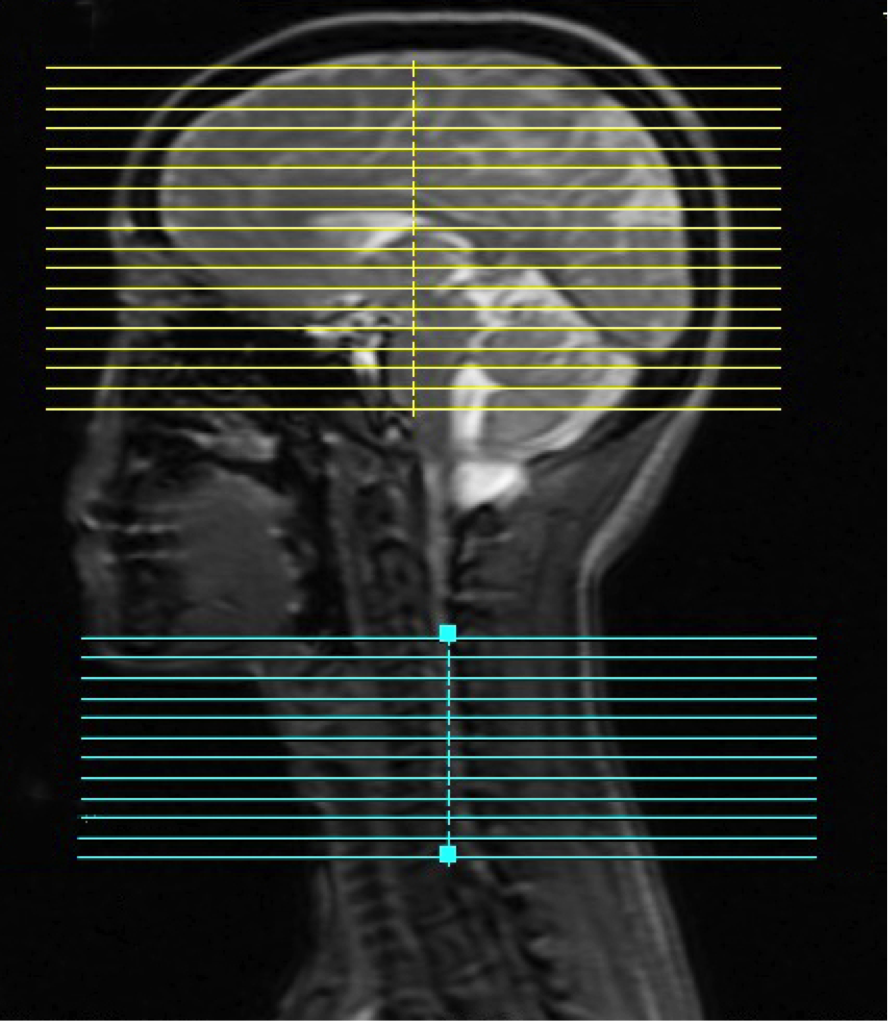
Seven healthy subjects performing a fist-clenching task were scanned in a GE 3T Discovery 750 using published parameters5. 18 brain and 12 spinal cord slices (spinal cord C5-C8) are acquired within the same TR (Fig.1). The fMRI task is a block design (15s on/off, 7.5 minutes duration). Subjects are instructed to clench both fists at 80% of maximum force during on-blocks. Physiological data is collected by respiratory belt and pulse oximeter.
Each data set is analyzed thrice: (1) no physiological correction, (2) RETROICOR correction, (3) RETROICOR followed by RVHRCOR correction. RETROICOR and RVHRCOR corrections are performed before pre-processing and analysis steps described in Ref.5. Activation volume in brain is defined: Z>2.3 having cluster size>10 voxels. For spinal cord, activation volume is defined: Z>1.6. Only voxels within brain and spinal cord are subject to tSNR and activation volume calculation.
Results
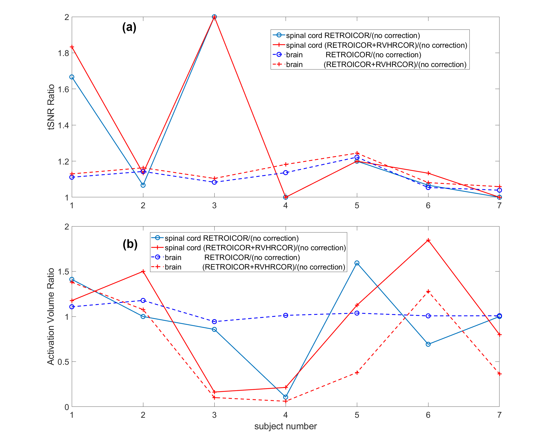
Figure 2a illustrates consistent increase in tSNR ratio: RETROICOR/(no correction) and (RETROICOR+RVHRCOR)/(no correction) calculated separately for brain and spinal cord. But there is significant variation, among subjects, in activation volume ratio due to these two corrections (Fig.2b).
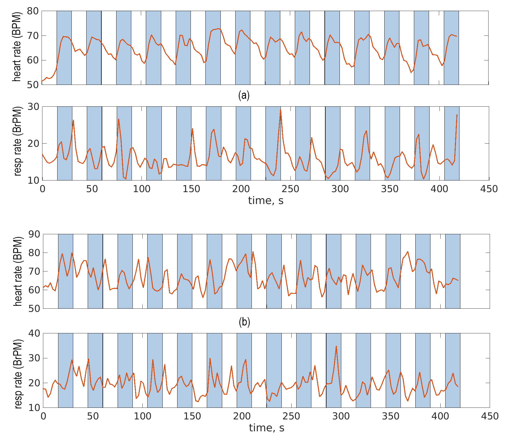
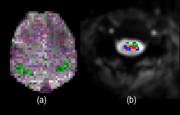
Subject 3 exhibits a significant drop in activation volume after applying RETROICOR+RVHRCOR; brain and spinal cord activation volume decreased by 90% and 84% respectively. This is explained by inspection of the physiological data which shows subject 3’s heart rate and breathing closely following task cycle as shown in Fig.3a. Fig.4a shows that RETROICOR+RVHRCOR remove false activation in brain vessels and gray matter not involved with task. While false activation in spinal cord is more difficult to discern, due to its small anatomy, it appears that RETROICOR+RVHRCOR erroneously remove both true and false activation (Fig.4b). Physiological noise removal using RETROICOR alone in spinal cord (blue) best reveals activation matching the anatomy.
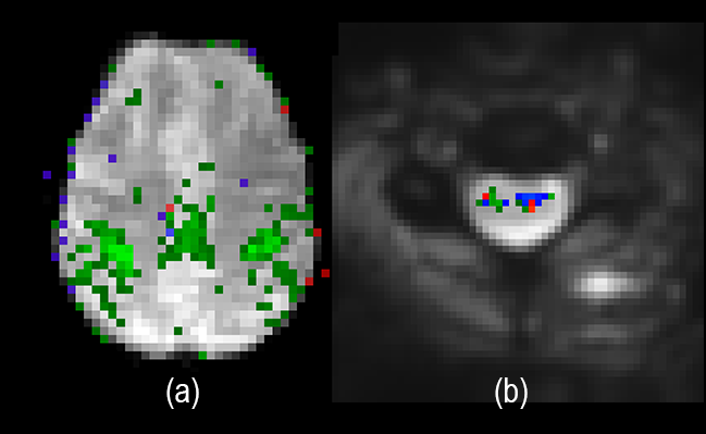
Subject 6 is at the other end of the spectrum, showing little correlation between cardiac/respiratory cycles and task (Fig.3b). After applying RETROICOR+RVHRCOR in brain and spinal cord, activation volume is respectively increased by 28% and 84% (Fig.1b). But there is a decrease in false activation along the edge of brain and an increase in activation in gray matter (Fig.5a). There is increased true activation in spinal cord localized at ventral horns (Fig.5b).
Conclusion
For a fist-clenching task, we have shown that RETROICOR and RVHRCOR consistently increase tSNR in both brain and spinal cord when compared with no physiological noise correction. On average, RETROICOR improves tSNR in brain and spinal cord by 11% and 29% respectively while RVHRCOR further improves tSNR in brain and spinal cord by 2% and 4% respectively (Fig.2a).Discussion
Although RETROICOR and RVHRCOR improve tSNR in both brain and spinal cord while removing false activation in brain, these corrections can remove true activation in spinal cord. These corrections do not always improve activation volume; even for the same subject within the same acquisition. RETROICOR removes cardiac and respiratory cyclic fluctuation that are expected common to brain and spinal cord. RVHRCOR does not reliably compensate long-term fluctuation of heart rate and respiratory volumes in spinal cord because its corresponding response function is derived from brain data. Before applying RETROICOR and RVHRCOR to data whose physiological noise may be correlated with task, examination of subject-level activation and noise is necessary because group analysis can average out individual variation. Examination with and without physiological correction is the only known way to determine whether subject-level activation were removed by noise correction. In general, an increase in activation volume after physiological noise correction is a good indicator of little correlation between physiology and task.Acknowledgements
NIH grants P41 EV0015891, K24 DA029262, T32 DA035165, General ElectricHealthcare, Gambhir-RSL Innovation Challenge Grant, and the Redlich Pain Endowment.
References
1. Glover, G. H., Li, T. Q. & Ress, D. Image-based method for retrospective correction of physiological motion effects in fMRI: RETROICOR. Magn. Reson. Med. 44, 162–167 (2000).
2. Chang, C., Cunningham, J. P. & Glover, G. H. Influence of heart rate on the BOLD signal: the cardiac response function. NeuroImage 44, 857–869 (2009).
3. Birn, R. M., Smith, M. A., Jones, T. B. & Bandettini, P. A. The Respiration Response Function: The temporal dynamics of fMRI signal fluctuations related to changes in respiration. NeuroImage 40, 644–654 (2008).
4. Kong, Y., Jenkinson, M., Andersson, J., Tracey, I. & Brooks, J. C. Assessment of physiological noise modelling methods for functional imaging of the spinal cord. Neuroimage 60, 1538–49 (2012).
5. Islam, H., Law, C. S. W., Weber, K. A., Mackey, S. C. & Glover, G. H. Dynamic per slice Shimming for Simultaneous Brain and Spinal Cord fMRI. Magn. Reson. Med. (2018). doi:10.1002/mrm.27388
Figures