3908
Recurrent Edge-Convolutional Neural Network for Individual Identification Using Resting-state fMRI Data1Department of Electrical and Computer Engineering, University of California, Riverside, Riverside, CA, United States, 2Department of Bioengineering, University of California, Riverside, Riverside, CA, United States, 3Center for Advanced Neuroimaging, University of California, Riverside, Riverside, CA, United States
Synopsis
Convolutional neural networks extract local features effectively on volumetric data. For the ROI-based fMRI data, we proposed the recurrent edge-convolutional neural network to model spatial coactivation pattern and dynamics. Edge-convolution could depict the relation between hyper-neighborhoods based on pre-computed functional connectivity and
INTRODUCTION
Individual variability in functional connectivity (FC) has been used as a connectomic fingerprinting to differentiate subjects1. However, traditional FC analysis focuses on static connectivity between brain networks or ROIs, which neglects the temporal changes or dynamics of connectivity. As a recent effort, recurrent neural network (RNN) was implemented to model spatiotemporal features of fMRI data for individual identification2. Though better performance was achieved, the initial RNN approach learns and updates spatial features using all ROIs simultaneously, ignoring the underlying functional and structural organization of the brain. In this work, we introduce the recurrent edge-convolutional architecture to model spatial coactivation pattern and dynamics of the fMRI data.METHODS
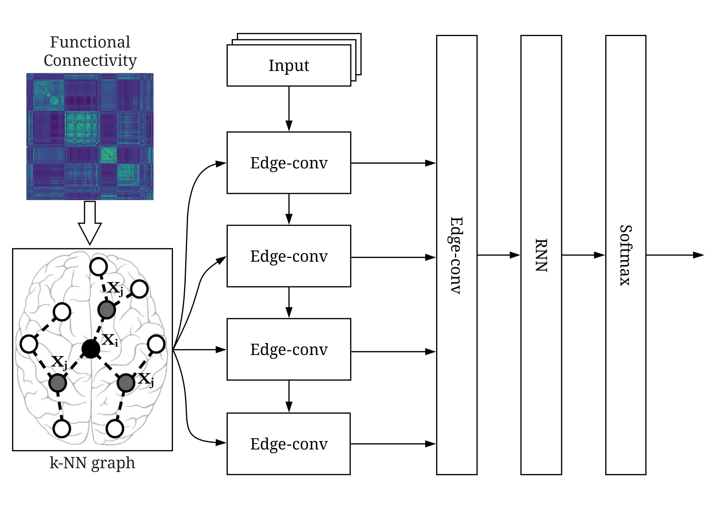
Traditional convolutional neural networks (CNNs) are effective in extracting spatial feature locally in Euclidean space on structured image grids. For the fMRI data analysis based on region-of-interests (ROIs), we introduced edge-convolution (Edge-conv) to extend convolutions on ROIs considering functional organization, as shown in Fig.1. Specifically, k-nearest neighbors (k-NN) were determined for each ROI to provide a neurophysiological-meaningful guidance for convolutions, where the FC was calculated by averaging across subjects in advance. The pointwise convolution was then performed within top-k neighbors with the asymmetric edge convolutional function h(xi,xj,θ)=hθ(xi,xj-xi), where hθ is the non-linear parametric function with learnable parameters θ, xi is the centroid and xj is one of the top-k neighbors3. Four Edge-conv layers were stacked with enlarged field-of-view for better capture of interactive activity. Another edge-conv layer took outputs from all preceding layers with feed-forward paths to alleviate the vanishing gradient problem. Lastly, a RNN layer was utilized to model temporal evolution, which was followed by a Softmax layer for the final classification.
We trained and tested our network on the resting-state fMRI data with 100 subjects from the Human Connectome Project (54 females, mean age = 29.1 ± 3.7, repetition time = 0.72 s)4. Two hundred and thirty-six ROIs with 5-mm radius over the cerebral cortex were selected5, where BOLD signals within each ROI were averaged as the corresponding ROI signal. Two scans on the first day were used for training and other two scans on the second day were used for validation and testing, respectively. Each scan has 1200 frames in total. During training and validation, the number of input frames was set to 100, resulting in 2400 training samples and 1200 validation samples. The final identification accuracy was reported as the average performance on the testing dataset with different number of frames as input.
RESULTS
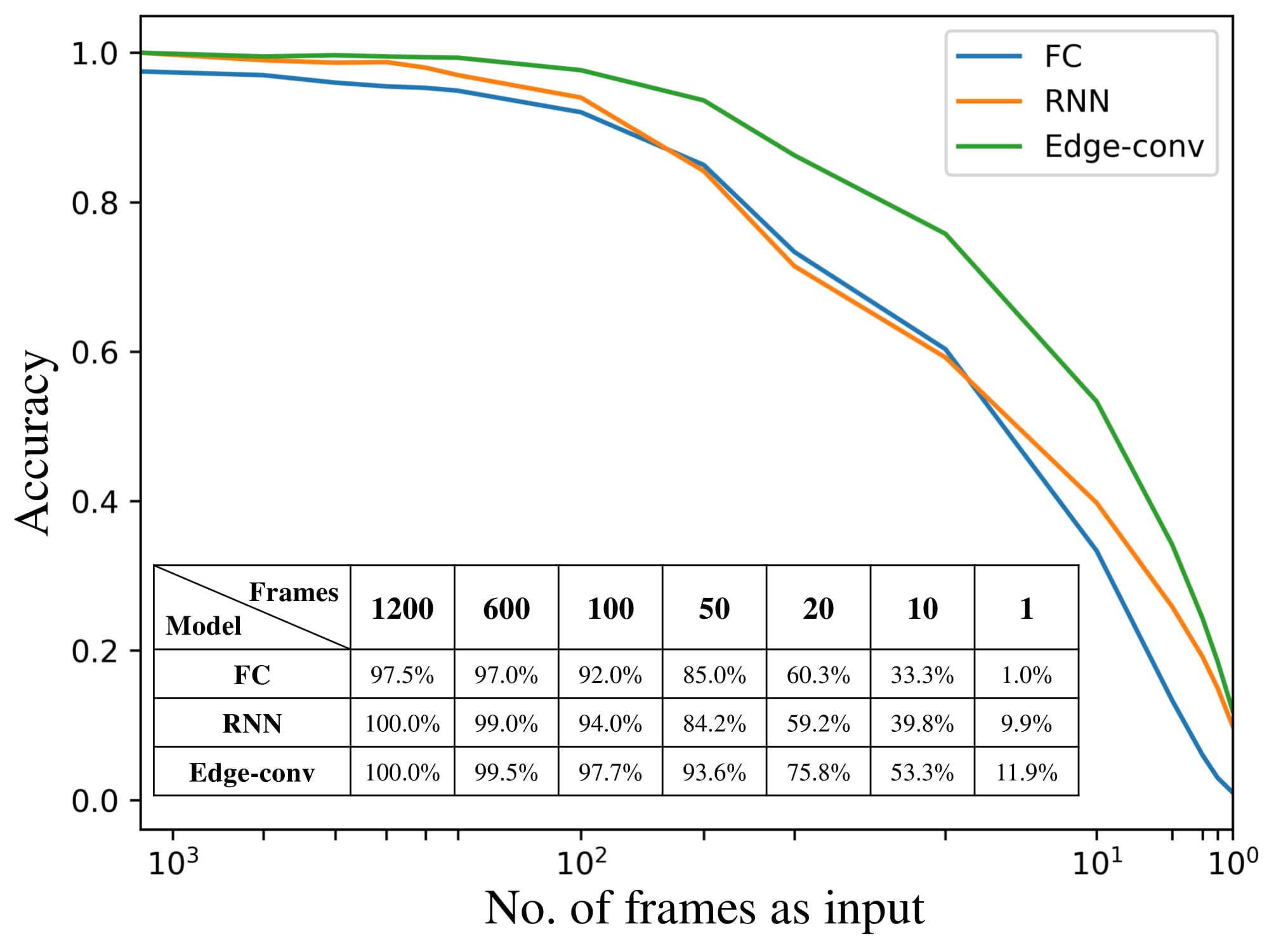
We compared our recurrent edge-convolutional approach with static FC1 and RNN2 using different number of frames, with the identification accuracy plotted and summarized in Fig.2. Our recurrent edge-convolutional model consistently achieved the highest accuracy, with over 2.5% performance improvement than FC on short clips of fMRI data (>100 frames). Especially for short clips (<100 frames), our recurrent edge-convolutional model could exceed FC and RNN by up to 20.8% and 16.6%. Under extreme situation with one single frame, our recurrent edge-convolutional model could still achieve 11.9% accuracy owing to the use of k-NN graph, when FC was totally not working.DISCUSSION
ROI-based fMRI data analysis could effectively reduce spatial complexity and highly reduce noise level of raw data on average. To extend convolution from grid-like data, we proposed edge-convolution on ROIs with the k-NN graph, where hyper-neighborhoods are decided for each ROI. Different from traditional CNNs extracting local patterns from close neighbors in Euclidean distance, our recurrent edge-convolutional architecture can accurately model coactivation from both local and remotely-connected neighbors defined by the k-NN graph. Experimental results showed that our approach always outperformed static FC and RNN model with better extraction of spatial interdependency. This is particularly true with limited number of frames, where much higher identification accuracy was achieved by effectively combining temporal information and spatial features from Edge-conv layers. While FC and RNN suffer from temporal variation, leading to unstable feature extraction and degraded identification performance.CONCLUSION
We introduced the edge-convolution for ROI-based fMRI data analysis. With the pre-computed k-NN graph, the neighborhood is determined for each ROI according to temporal correlation instead of the Euclidean metric. The coactivation between ROIs can be accurately depicted compared to traditional convolutions on raw data in volumetric representation. Experiment results demonstrated that our recurrent edge-convolutional model was able to achieve higher identification accuracy with spatiotemporal features extracted from resting-state fMRI data.Acknowledgements
No acknowledgement found.References
1 Finn E S, Shen X, Scheinost D, et al. Functional Connectome Fingerprinting: Identifying Individuals Using Patterns of Brain Connectivity. Nature neuroscience. 2015. 18: 1664.
2 Chen S, and Hu X P. Individual Identification Using Functional Brain Fingerprint Detected by Recurrent Neural Network. Brain connectivity. 2018.
3 Wang Y, Sun Y, Liu Z, et al. Dynamic Graph Cnn for Learning on Point Clouds. arXiv preprint arXiv:1801.07829. 2018.
4 Van Essen D C, Smith S M, Barch D M, et al. The Wu-Minn Human Connectome Project: An Overview. Neuroimage. 2013. 80: 62-79.
5 Power J D, Cohen A L, Nelson S M, et al. Functional Network Organization of the Human Brain. Neuron. 2011. 72: 665-678.
Figures