3903
Effect of meditation on brain function activated by an attention task1Computer Science, Binghamton University, Binghamton, NY, United States, 2Cornell University, Ithaca, NY, United States
Synopsis
The effect of meditation on brain functional activation when engaged in an attention task was evaluated longitudinally using BOLD fMRI in nine young healthy subjects. Functional activation before and after meditation practice was compared and the change of functional activation was correlated with practice time. Following the meditation practice, response time to the attention task was significantly shorter; functional activation in the somatomotor network, frontal, basal ganglia, and insular regions was significantly reduced; more practice time was associated with less reduced activation. The findings suggest that meditation can improve brain efficiency but may have a complex neural mechanism.
Introduction
Focused meditation has been shown to impact attention [1-3], emotional regulation [4-6], and thinking patterns [7-9]. The functional neuroimaging studies found brain activation differences between experienced meditators and nonmeditators when engaged in attention tasks [2, 3] and emotional regulation tasks [5, 10]. To understand the causal relationship between meditation and brain functional change, a longitudinal design of the study is preferred. We aim to evaluate the effects of 2-month meditation on novice meditators when engaged in an attention task.
Methods
Nine young healthy participants (age: 19.22 ± 0.44 years old, 3 females, recruited from a university meditation class) were imaged using a GE 3T MR 750 scanner. Subjects were instructed to practice focused meditation for a minimum of 10 minutes each time and at least 5 times per week. They were allowed to choose their own focus: their breath, a point on the wall, a phrase, or anything else as they saw fit. Subjects received 3D T1-weighted Magnetization Prepared RApid Gradient Echo (MPRAGE) images for image registration and task-related blood oxygenation level-dependent (BOLD) functional MRI (fMRI) sequence with visual oddball paradigm for measurement of brain activation. Subjects were scanned with the same protocol at the baseline and after 2-month meditation practice (practice duration: 69.44 ± 4.98 days, practice time: 557.78 ± 490.78 minutes). fMRI visual stimuli, ‘X’ and ‘O’ were randomly shown (the frequency ratio set as 20:80) on an LCD monitor viewed through a mirror mounted on the MRI scanner’s head coil. The inter-stimulus interval (ISI) was ranging from 4s to 5s and stimulus duration was set as 500 milliseconds. Subjects were asked to press “left button” for ‘X’ and “right button” for ‘O’. During the remaining time (3.5s to 4.5s), the subjects were asked to focus on the fixation point. BOLD functional images (TR = 2s) were obtained using T2* weighted echo planar imaging (EPI) sequence.
BOLD images were performed with slice timing correction, motion correction, normalization to the standard MNI space using T1 weighted image as intermediate, and spatial smoothing (FWHM=6mm). The individual activation maps were produced using the SPM8 general linear model with the onset times of ‘X’ and ‘O’ and their temporal derivatives as regressors. The individual contrast images, ‘X’, ‘O’, ‘X+O’, and ‘X-O”, were generated for each subject and each condition (baseline or follow-up). The individual contrast images were compared between baseline and follow-up using paired t tests. The general linear model (GLM) was used to detect the potential association between practice time and the change of contrast images with gender as a covariate. The statistical maps were thresholded using a voxel-level p value of 0.02/0.01. A cluster-level p value of 0.05 was used to correct for multiple comparisons.
Results
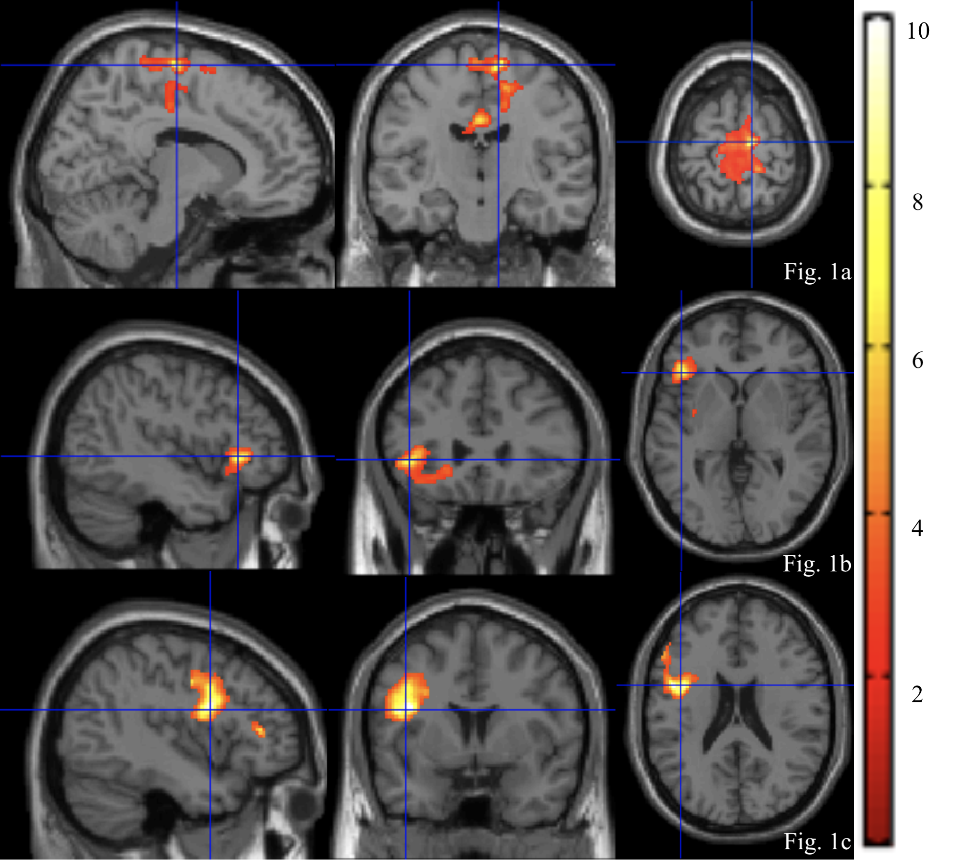
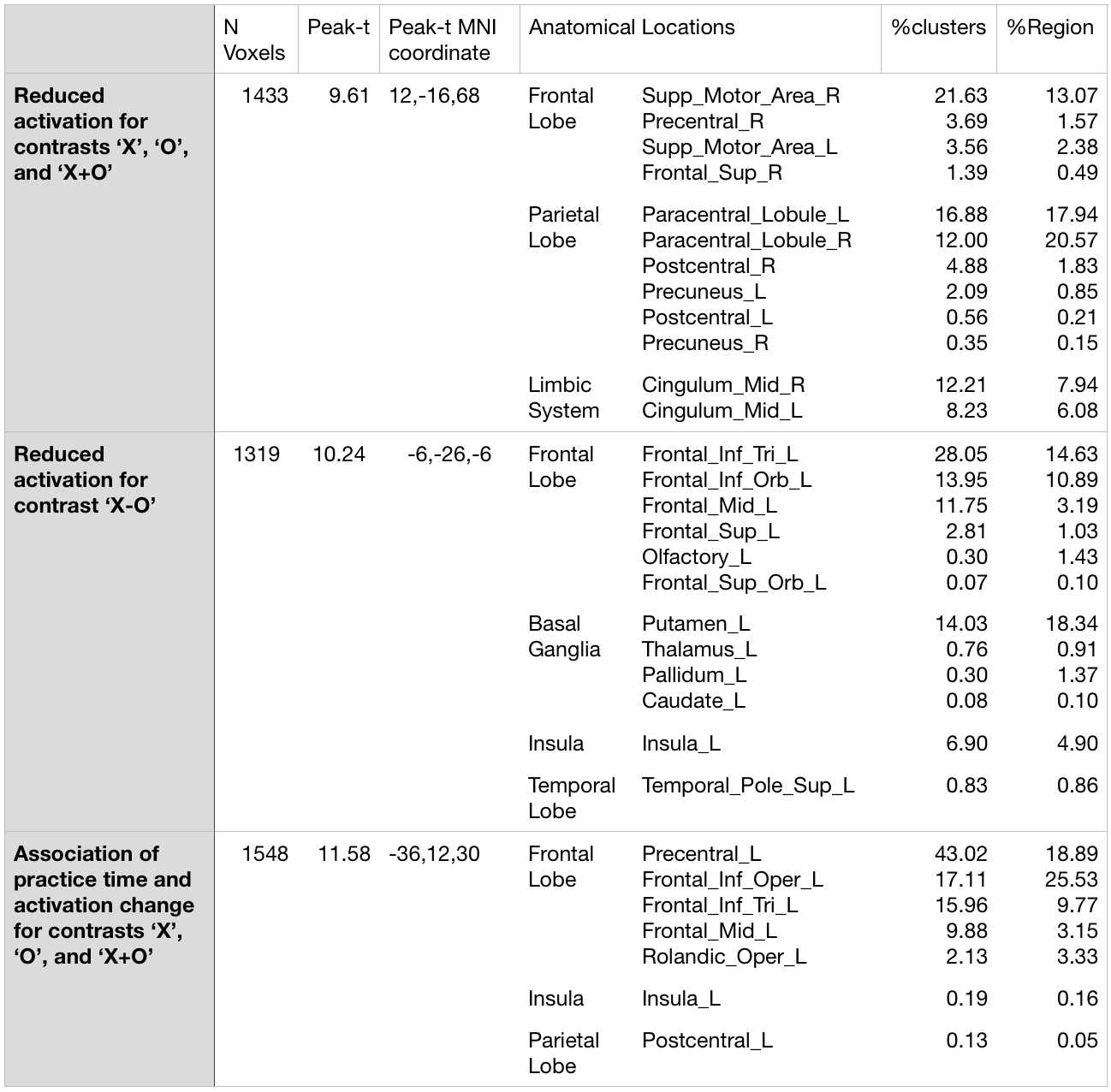
Response time to ‘X’ and ‘O’ was significantly shorter (baseline: 356.73 ± 19.73 ms, follow-up: 325.67 ± 30.35 ms, p = 0.050) but percentage of correct response was not significantly different after meditation practice. After 2-month meditation practice, significant reduced activation (voxel-level p value of 0.02) was found in the somatomotor network regions (Fig. 1a and Table 1), including bilateral supplementary motor and paracentral regions for ‘X’, ‘O’ and ‘X+O’ contrasts, and in the left frontal, basal ganglia, and insular regions for ‘X-O’ contrast (Fig. 1b and Table 1). We also found the positive association between practice time and the change of activation for ‘X’, ‘O’ and ‘X+O’ contrasts in the frontal regions (Fig. 1c and Table 1).
Discussion
Following 2-month meditation practice, novice meditators had reduced activation in the somatomotor network region and the left frontal, basal ganglia, and insular regions in an attention task. These results are in a good agreement with the decreased nodal strength in the somatomotor network region after 8-week meditation training in an elderly cohort [11] and decreased activation in the regions after a 7-day meditation with Stroop Word-Color Task (SWCT), which also requires attention control [12]. The findings suggest that meditation improves brain efficiency with less functional activation in attention-related regions to achieve the same task performance. However, the association between more practice time and reduced brain activation may indicate the effect of meditation is not linear, which supports the potential “U” shape hypothesis of meditation practice [13]. This study provides further support of short-term meditation as a potentially beneficial method for efficient attention control.Acknowledgements
This research was supported by the State University of New York at Binghamton.References
1. Goldin, P., et al., MBSR vs aerobic exercise in social anxiety: fMRI of emotion regulation of negative self-beliefs. Soc Cogn Affect Neurosci, 2013. 8(1): p. 65-72.
2. Kozasa, E.H., et al., Meditation training increases brain efficiency in an attention task. Neuroimage, 2012. 59(1): p. 745-9.
3. Pagnoni, G., Dynamical properties of BOLD activity from the ventral posteromedial cortex associated with meditation and attentional skills. J Neurosci, 2012. 32(15): p. 5242-9.
4. Farb, N.A., et al., Minding one's emotions: mindfulness training alters the neural expression of sadness. Emotion, 2010. 10(1): p. 25-33.
5. Taylor, V.A., et al., Impact of mindfulness on the neural responses to emotional pictures in experienced and beginner meditators. Neuroimage, 2011. 57(4): p. 1524-33.
6. Grimm, S., et al., Altered negative BOLD responses in the default-mode network during emotion processing in depressed subjects. Neuropsychopharmacology, 2009. 34(4): p. 932-43.
7. Northoff, G. and F. Bermpohl, Cortical midline structures and the self. Trends Cogn Sci, 2004. 8(3): p. 102-7.
8. McGuire, P.K., et al., Brain activity during stimulus independent thought. Neuroreport, 1996. 7(13): p. 2095-9.
9. Northoff, G., et al., Self-referential processing in our brain--a meta-analysis of imaging studies on the self. Neuroimage, 2006. 31(1): p. 440-57.
10. Goldin, P.R. and J.J. Gross, Effects of mindfulness-based stress reduction (MBSR) on emotion regulation in social anxiety disorder. Emotion, 2010. 10(1): p. 83-91.
11. Cotier, F.A., R. Zhang, and T.M.C. Lee, A longitudinal study of the effect of short-term meditation training on functional network organization of the aging brain. Sci Rep, 2017. 7(1): p. 598.
12. Kozasa, E.H., et al., Effects of a 7-Day Meditation Retreat on the Brain Function of Meditators and Non-Meditators During an Attention Task. Front Hum Neurosci, 2018. 12: p. 222.
13. Brefczynski-Lewis, J.A., et al., Neural correlates of attentional expertise in long-term meditation practitioners. Proc Natl Acad Sci U S A, 2007. 104(27): p. 11483-8.
Figures