3900
Neural signatures underlying cognitive styles of field independence and field dependence: Evidence from resting-state fMRI1Institute of Nuclear Medicine and Allied Sciences (INMAS), Delhi, India, 2Defence Institute of Psychological Research (DIPR), Delhi, India
Synopsis
Cognitive style refers to the individual differences in the distinct preferences to think, learn, solve problems and to perceive and organize information about the surrounding space. Field dependence/ independence (FDI) is the most widely studied cognitive style and is measured by Group Embedded Figures Test (GEFT) that requires a participant to locate the simple shape embedded in a complex figure. FI subjects are less influenced by the information from the prevailing visual fields and perform better on GEFT as compared to the FD subjects. The cognitive style of an individual has been shown to be related to their cognitive functioning especially spatial memory performance, learning and retrieval of
Purpose
Cognitive style refers to the individual differences in the distinct preferences to think, learn, solve problems and to perceive and organize information about the surrounding space.1,2 Field dependence/ independence (FDI) is the most widely studied cognitive style and is measured by Group Embedded Figures Test (GEFT) that requires a participant to locate the simple shape embedded in a complex figure.3 FI subjects are less influenced by the information from the prevailing visual fields and perform better on GEFT as compared to the FD subjects. The cognitive style of an individual has been shown to be related to their cognitive functioning especially spatial memory performance, learning and retrieval of navigational environment, personality characteristic and social behaviour.1,2,4 However, brain-behavior relationship of FDI cognitive styles has been little investigated.2 The present study aimed to assess the underlying resting state connectivity networks in relation to the FDI cognitive styles measured by GEFT.Methods
Thirty seven right-handed healthy and educated (graduates/ post graduates) subjects (male – 14, female – 23, mean age –21.75 years, SD – 2.03 years) participated in the study. The study was carried out using 3T whole body MR system (Magnetom Skyra, Siemens, Germany) with a circularly polarized 20 channel matrix head and neck coil and 45 mT/m actively shielded gradient system. Functional brain volumes were acquired using echo-planar T2* -weighted imaging sequence (TE = 30 ms, TR = 2000 ms, FOV = 240 mm, flip angle = 90°, voxel size = 3.75X3.75X5 mm3, 30 interleaved 5-mm thick slices without inter-slice gap per brain volume). Total scanning time was 410 seconds (205 brain volumes), during which the subjects were asked to keep their eyes closed without thinking of anything in particular and not falling asleep. The rs-fMRI data was analyzed using Multivariate Exploratory Linear Optimized Decomposition into Independent Components (MELODIC) toolbox of FSL (FMRIB's Software Library, www.fmrib. ox.ac.uk/fsl). General Linear Model was defined to create multi-subject design matrix defining groups (two groups: high GEFT (HEFT) and low GEFT (LEFT); median split on the basis of participants’ GEFT scores) and contrast files (two contrasts: HEFT vs LEFT and LEFT vs HEFT). A regressor for age (mean centered) was also added. Dual regression approach was then applied on 11 RSNs that were identified.5 Masks were created for all the eleven resting state networks. Voxel-wise analyses of the group differences in different networks between the HEFT and LEFT groups were carried out using FSL randomize non-parametric permutation-testing with 10000 permutations function per contrast. Threshold-Free Cluster Enhancement6 was used to control for multiple comparisons (significance threshold p < 0.05 FWE corrected). The statistical maps were then upsampled to standard MNI 1 mm brain Montreal atlas to better localize the areas of RSNs modifications. The Harvard-Oxford cortical and subcortical atlases were used to identify the anatomical characteristics of the resulting Probabilistic Independent Component Analyses (PICA) maps using ‘atlasquery’ tool. A similar approach was used for a correlation analysis between participants’ GEFT scores and the functional connectivity maps.Results
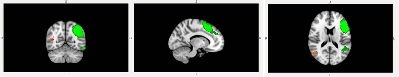
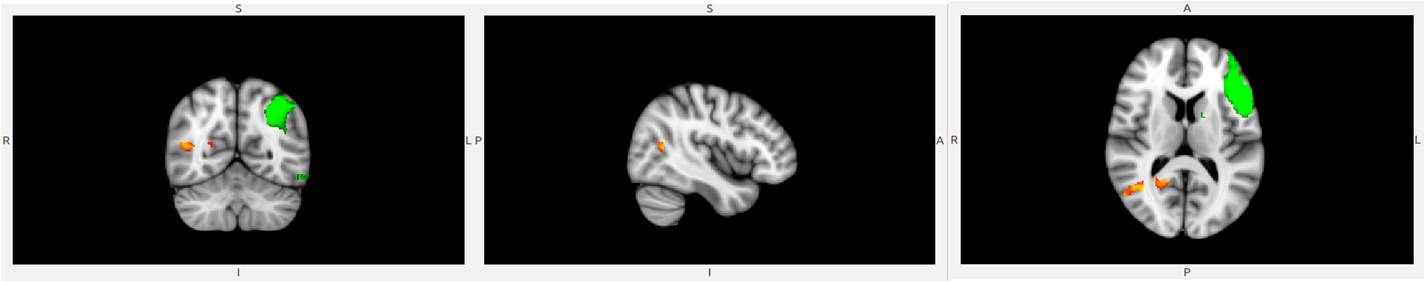
Subjects with GEFT scores < 12 were considered as LEFT group (male – 4, female – 14) and those with score > 12 were considered as HEFT group (male – 10, female – 9). Identified RSNs from group MELODIC output included executive network (EN), fronto-parieto-temporal network (FPTN), dorsal attention network (DAN), default mode network (DMN); anterior DMN (ADMN); auditory network (AN); somato-motor network (SMN); medial visual network (MVN); lateral visual network (LVN); right fronto-parietal attention network (RFPN); left fronto-parietal attention network (LFPN). HEFT subjects showed significantly increased functional connectivity of the right hemispheric occipito-temporal regions such as lateral occipital cortex, precuneous cortex, intracalcarine cortex, cuneal cortex, middle temporal gyrus (temporo-occipital part) with the LFPN (Figure 1). Significantly positive correlation was also obtained between the GEFT scores and the functional connectivity of the right hemispheric occipito-temporal regions with the LFPN (Figure 2).Discussion
The frontoparietal attention network is anatomically and functionally interposed between the DAN and DMN and underpins attention, visuospatial working memory, and executive control functions. The network includes both the dorsal attention network and ventral attention network. The right hemispheric regions showing increased connectivity with LFPN are involved in visual processing, object recognition and perception, mental imagery and memory processes. GEFT assesses visuospatial tendency indicative of spatial orienting or global-local processing.2 A greater resting state coherence between the occipito-temporal regions of the right hemisphere with the left fronto-parietal attention network in individuals with high GEFT scores might underlie superior local visual processing for FI individuals.Conclusion
Preliminary findings thus contribute towards understanding the brain-behaviour relationship associated with cognitive style of FDI.Acknowledgements
This work was supported by DRDO Project No. S&T-14/DIPR-734.References
1. Tascón L, Boccia M, Piccardi L, et al. Differences in Spatial Memory Recognition Due to Cognitive Style. Front Pharmacol. 2017; 8: 550.
2. Hao X, Wang K, Li W. Individual Differences in Brain Structure and Resting Brain Function Underlie Cognitive Styles: Evidence from the Embedded Figures Test. PLOS ONE. 2013; 8(12): e78089.
3. Witkin HA, Oltman PK, Raskin E, et al. A manual for the Group Embedded Figures Test. Menlo Park, CA: Mind Garden, Inc, 1971.
4. Boccia M, Vecchione F, Piccardi L. Effect of Cognitive Style on Learning and Retrieval of Navigational Environments. Front Pharmacol. 2017; 8: 496.
5. Beckmann CF, DeLuca M, Devlin JT. Investigations into resting-state connectivity using independent component analysis. Philos. Trans. R. Soc. Lond B Biol Sci. 2005; 360: 1001–1013.
6. Smith SM, Nichols TE. Threshold-free cluster enhancement: addressing problems of smoothing, threshold dependence and localisation in cluster inference. Neuroimage 2009; 44: 83–98.
Figures