3899
Altered the spinal trigeminal nucleus spontaneous brain activity and functional connectivity in female migraineurs without aura1East China Normal university, Shanghai, China, 2Shanghai Jiao Tong University School of Medicine, Shanghai, China
Synopsis
The purpose of this study was to investigate spontaneous neuron activity in spinal trigeminal nucleus (sTN) and its intrinsic functional connectivity (FC) with other key structures in migraineurs during resting-state fMRI scans. Thirty-five female migraine without aura (MWoA) during headache interictal phase and 35 age matched female HC were recruited. We found higher ALFF z-value of left sTN in female MWoA. We also found significantly weaker FC between the sTN and caudate putamen (CPu), and dorsolateral prefrontal cortex (DLPFC). Our results help us better understand the pathology of migraine.
Introduction
Spinal trigeminal nucleus (sTN) have been identified as a conduction site enrolled in the ascending pain pathways and descending pain modulation system1. Previous studies have demonstrated functional and structural abnormality of sTN in migraineurs. However, the spontaneous brain activity of sTN and its intrinsic functional connectivity (FC) with other brain areas in migraineurs remains unclear. We proposed that altered neuron activity in sTN and its altered functional coupling with other key structures may result in a dysfunction of pain processing and modulation.Materials and Methods:
Thirty-five female migraineurs without aura (MWoA) (mean ± SD age=41.1±12.2 years) and 35 female healthy controls (HC) (mean ± SD age = 41.6±12.1 years) were enrolled in our study. Rest-state fMRI images were acquired using an EPI sequence with the following parameters: TR/ TE = 2000/ 30 ms, 33 slices, 210 volumes. Structural scans were acquired for registration and normalization of the functional images by using a 3-dimensional magnetization-prepared rapid-acquisition gradient-echo pulse sequence (TR/TE = 2530/ 2.34 ms, 192 slices). Seed MNI coordinates: left sTN = -3;-40;-54; right sTN = 3;-40;-54, reported from Schulte, Allers et al. 20182. Seed regions were 3mm spherical regions. We calculated ALFF (0.01~0.1Hz) and seed based FC map using Data Processing Assistant for Resting-State fMRI software. Two-sample t-test was conducted to compare ALFF z-value in the sTN between female MWoA and HC. The seed based FC maps between female MWoA and the HC were compared using voxel-wise two-sample T-tests in spm12 to detect brain network difference.Results:
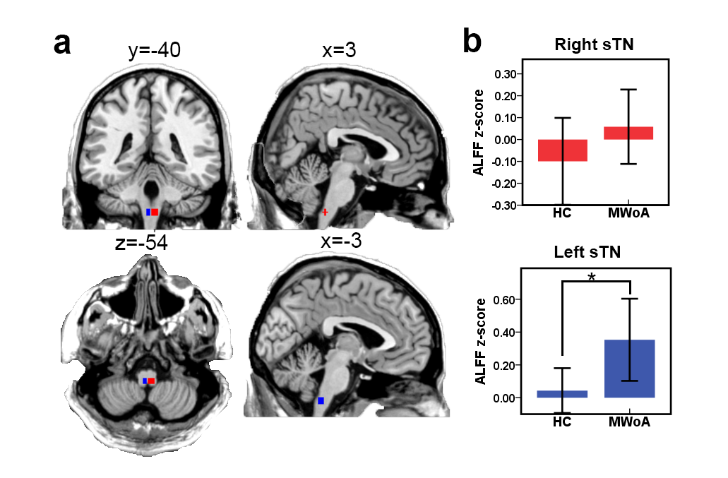
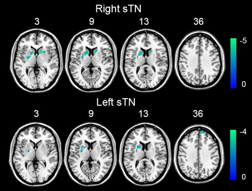
Compared with controls, The ALFF z-value of left sTN of female MWoA is significantly higher than controls’ (Fig.1). The female MWoA showed significantly weaker FC between the right sTN and the bilateral caudate putamen (Fig. 2). Moreover, female MWoA also showed significantly weaker FC between left sTN and left putamen and right superior frontal gyrus (Fig. 2)Discussion:
We found increased spontaneous activity of the sTN. STN have been identified as a conduction site enrolled in the ascending pain pathways and descending pain modulation system1. Higher spontaneous activity in sTN may reflect an abnormal state of sTN in the spinal and trigeminal nociceptive pathways. Our results attach vital importance to sTN in the pathology of migraine. The female MWoA showed significantly weaker FC between the right sTN and the bilateral caudate putamen and significantly weaker FC between left sTN and left putamen and right superior frontal gyrus. Our results suggested the atypical connectivity of sTN to CPu and DLPFC were associated with impaired pain processing and modulatory processes in female MWoA, which could contribute to the pathology of migraine.Conclusion:
Our results showed increased evidence that sTN is involved in the pathophysiological migraine. The weaker FC of sTN to CPu and DLPFC may result in deficits of pain modulation. Our results help us better understand the pathology of migraine.Acknowledgements
This work was supported by grants from the National Natural Science Foundation of China (Nos. 81571658 to X. X. Du).References
1. Goadsby PJ, Holland PR, Martins-Oliveira M, Hoffmann J, Schankin C and Akerman S. Pathophysiology of migraine: a disorder of sensory processing. Physiological reviews. 2017; 97: 553-622.
2.Schulte LH, Allers A and May A. Visual stimulation leads to activation of the nociceptive trigeminal nucleus in chronic migraine. Neurology. 2018; 90: e1973-e8.
Figures