3898
Hippocampal atrophy and functional connectivity disruption in cirrhotic patients with minimal hepatic encephalopathy1Fujian Medical University Union Hospital, Fuzhou, China, 2Siemens Healthcare, Fuzhou, China
Synopsis
The hippocampus is a crucial pathological node for minimal hepatic encephalopathy (MHE) and it is associated with various cognitive impairments. We assessed hippocampal FC alterations in relation to regional volume and their associations with PHES results of cirrhotic patient and reached the following findings. (i) Hippocampal volume is significantly lower in MHE. (ii) A disruption of hippocampal connectivity to the default-mode network was observed in the MHE patients. (iii) The bilateral hippocampal FC strength (but not hippocampal volume) was positively correlated with the PHES results of the cirrhotic patients and aggravate with HE progression. Our findings revealed that both structural and functional abnormalities of the hippocampus are involved in the mechanism underlying cognitive deterioration with disease progression.
Purpose
The hippocampus is a crucial pathological node for minimal hepatic encephalopathy(MHE) and it is associated with various cognitive impairments. Investigations on alterations involving hippocampal morphology and functional connectivity (FC) in MHE are limited. This study aimed to simultaneously evaluate hippocampal volume and FC alterations and their association with cognitive decline in MHE.Materials and Methods
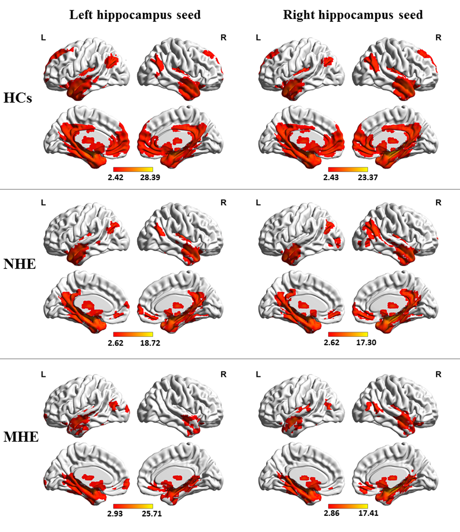
Data were collected on a MAGNETOM Prisma 3.0T scanner (Siemens Healthcare, Erlangen, Germany) with a 64-channel head coil. Twenty-two cirrhotic patients with MHE, 31 cirrhotic patients without MHE (NHE), and 43 healthy controls underwent high-resolution T1-weighted imaging, resting-state functional magnetic resonance imaging, and cognition assessment based on Psychometric Hepatic Encephalopathy Score (PHES). The structural images were preprocessed using a voxel-based morphometry method, during which hippocampal volume was measured. The hippocampal connectivity network was identified using seed-based correlation analysis. Hippocampal volume and FC strength were compared across the three groups and correlated against the PHES results of the cirrhotic patients.Results
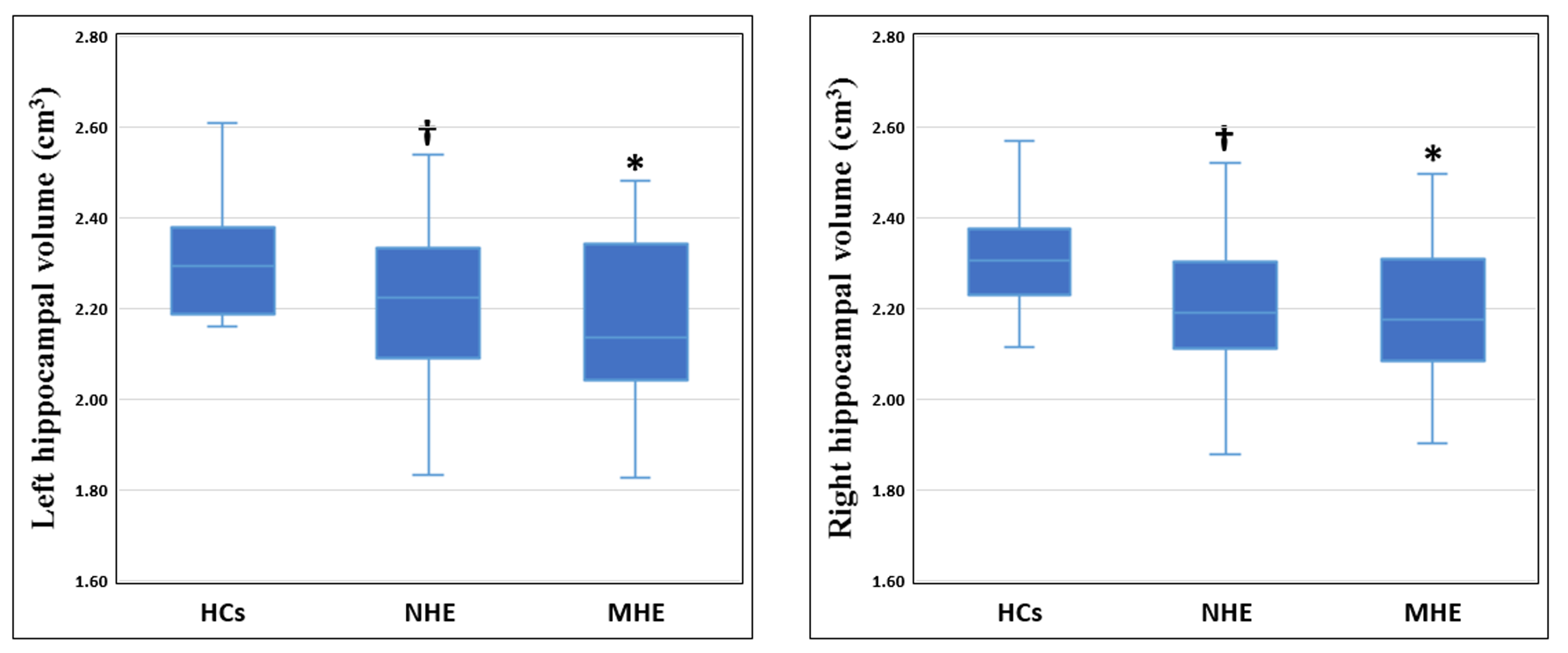
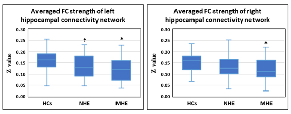
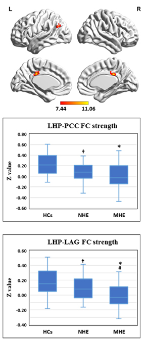
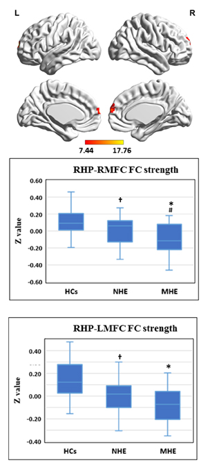
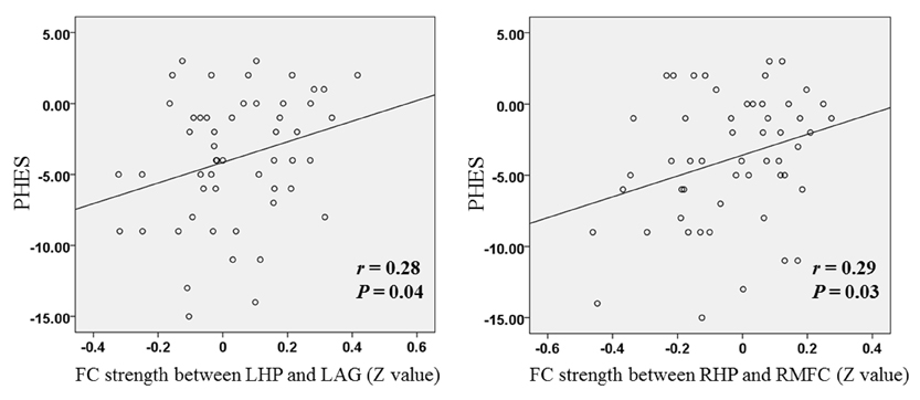
Compared to the controls, MHE patients exhibited a significantly lower bilateral hippocampal volume. A slight decrease in hippocampal volume was obtained from NHE to MHE, but it did not reach statistically significance. In addition, the average FC strength of the bilateral hippocampal connectivity network was significantly lower in the MHE patients. In particular, the MHE patients showed a decrease in FC involving the left hippocampus to bilateral posterior cingulate gyrus and left angular gyrus. The MHE patients also showed FC reduction between the right hippocampus and bilateral medial frontal cortex. A progressive reduction in hippocampal FC from NHE to MHE was also observed. The bilateral hippocampal FC strength (but not hippocampal volume) was positively correlated with the PHES results of the cirrhotic patients.Conclusion
Our assessment of MHE patients revealed decreased hippocampal volume, which suggests regional atrophy, and reduced hippocampal connectivity with regions that are primarily involved in the default-mode network, thereby suggesting a functional disconnection syndrome. These alterations reveal the mechanisms underlying cognitive deterioration with disease progression.Acknowledgements
This study was funded by the grants from the National Natural Science Foundation of China (No.81501450), Fujian Provincial Science Fund for Distinguished Young Scholars (No. 2018J06023), Fujian Provincial Program for Distinguished Young Scholars (No.2017B023), and Fujian Provincial Health Commission Project for Scientific Research Talents (2018-ZQN-28).References
1.Agrawal, S., Umapathy, S., Dhiman, R.K., (2015). Minimal hepatic encephalopathy impairsquality of life. J Clin Exp Hepatol 5, S42-48.10.1016/j.jceh.2014.11.006
2.Bird, C.M., (2017). The role of the hippocampus in recognition memory. Cortex 93,155-165.10.1016/j.cortex.2017.05.016
3.Blankenship, S.L., Redcay, E., Dougherty, L.R., Riggins, T., (2017). Development ofhippocampal functional connectivity during childhood. Human Brain Mapping 38,182-201.10.1002/hbm.23353
4.Brueggen, K., Kasper, E., Dyrba, M., Bruno, D., Pomara, N., Ewers, M., Duering, M., Burger,K., Teipel, S.J., (2016). The Primacy Effect in Amnestic Mild Cognitive Impairment:Associations with Hippocampal Functional Connectivity. Front Aging Neurosci 8,244.10.3389/fnagi.2016.00244
5.Lisman, J., Buzsaki, G., Eichenbaum, H., Nadel, L., Ranganath, C., Redish, A.D., (2017).Viewpoints: how the hippocampus contributes to memory, navigation and cognition. NatNeurosci 20, 1434-1447.10.1038/nn.4661
6.Qi, R., Zhang, L.J., Zhong, J., Zhang, Z., Ni, L., Zheng, G., Lu, G.M., (2013b). Disruptedthalamic resting-state functional connectivity in patients with minimal hepatic encephalopathy.Eur J Radiol 82, 850-856.10.1016/j.ejrad.2012.12.016S0720-048X(12)00644-4 [pii]
7.Zarei, M., Beckmann, C.F., Binnewijzend, M.A., Schoonheim, M.M., Oghabian, M.A.,Sanz-Arigita, E.J., Scheltens, P., Matthews, P.M., Barkhof, F., (2013). Functional segmentationof the hippocampus in the healthy human brain and in Alzheimer's disease. Neuroimage 66,28-35.10.1016/j.neuroimage.2012.10.071
8.Zeidman, P., Maguire, E.A., (2016). Anterior hippocampus: the anatomy of perception,imagination and episodic memory. Nature Reviews Neuroscience 17,173-182.10.1038/nrn.2015.24
9.Zhang, G., Cheng, Y., Liu, B., (2017). Abnormalities of voxel-based whole-brain functionalconnectivity patterns predict the progression of hepatic encephalopathy. Brain Imaging Behav11, 784-796.10.1007/s11682-016-9553-210.1007/s11682-016-9553-2 [pii]
Figures