3894
The effect of 2-month meditation on brain functional connectivity1Computer Science, State University of New York at Binghamton, Binghamton, NY, United States, 2Cornell University, Ithaca, NY, United States
Synopsis
The sustained effect of meditation on brain functional connectivity (FC) was evaluated longitudinally in ten college students. FC maps before and after meditation practice were compared and the changes of FC were correlated to practice time. We observed a significantly increased FC between the default mode network (DMN) and the dorsal attention network (DAN), and within DAN. The increased FC between DMN and DAN and within DAN was associated with increased practice time. The findings support fast shifting of attention after practicing meditation for two months and lay a foundation for its clinical transformation if confirmed in a larger study.
Introduction
Focused meditation has been shown to influence human brain networks, especially the default mode network (DMN) [1-4] and attention networks [5-7]. Most of these studies were performed using traditional fMRI techniques with single-echo echo-planar imaging (EPI) acquisition and mainly concentrated on the functional connectivity (FC) difference of brain networks between experienced meditators and nonmeditators during meditation. It is interesting to ask whether the changes in brain networks can be sustained during the rest period, even when meditation is not being performed, for its potential benefits in a clinical population. Here, we used a longitudinal design to explore the sustained change in brain FC during rest following 2-month meditation practice. Resting-state fMRI technique with multi-echo EPI [8] was used for data acquisition due to its effective removal of scanner drifts, motion artifacts, cardiac pulsations, and respiratory motions.Methods
All studies were conducted at the Cornell University MR Facility using a GE 3T MR 750. Volunteers received 3D T1-weighted Magnetization Prepared RApid Gradient Echo (MPRAGE) images for image registration and resting-state multi-echo EPI sequence [8, 9] for FC measurements before and after 2-month meditation practice (practice duration: 66.50 ± 4.14 days, practice time: 574.00 ± 312.30 minutes). Ten college students (19.20 ± 0.28 years old, age range: 19 to 20 years old, 4 females) were recruited from a university meditation class. Subjects were instructed to practice focused meditation for a minimum of 10 minutes each time and at least 5 times per week. They were allowed to choose their own focus: their breath, a point on the wall, a phrase, or anything else as they saw fit. At the end of follow-up scans, volunteers reported their practice time.
For each subject, multi-echo BOLD signal time series were transformed to the standard brain space using the T1-weighted images as an intermediate, and the neural-related BOLD signal time series was derived by removing the non-BOLD related artifacts with independent component analysis (ICA) and TE-dependence analysis [8, 10]. Five seed regions of interest [11] were chosen from DMN and attention networks: the posterior cingulate cortex(PCC), the left visual area (LMT), the right visual area (RMT), the left superior parietal area (LSPL), and the right superior parietal area (RSPL). LSPL and RSPL ROIs are located in the dorsal attention network (DAN). Individual FC maps were calculated from artifact-cleaned BOLD signal time series and also from traditional BOLD signal time series (averaged across multiple echo without artifact removal) using Pearson correlation between time series from the seed ROIs and those from individual voxels throughout the entire brain. Individual FC maps were transformed into z score maps by using a Fisher z transformation to improve normality for group level t tests. The z score maps were smoothed with a 6×6×6 mm Gaussian kernel. The z score maps before and after meditation practice were compared using SPM8 via a paired t test with gender as a covariate. Age was not considered as a covariate because the maximum difference among ages of our subject is 1. The difference of the z score maps before and after practice meditation was modeled using SPM8 via multiple linear regression with gender and practice time as covariates. The statistical maps were thresholded using a voxel-level p value of 0.01. A cluster-level p value of 0.05 was used to correct for multiple comparisons.
Results & Discussion
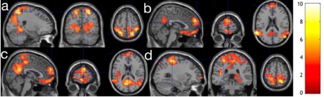
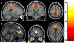
Without artifact removal from the multi-echo BOLD technique, no difference of FC with any seed ROI was observed between baseline and follow-up FC images. After artifact removal using the multi-echo BOLD technique, we observed significantly increased FC between the PCC seed and dorsal attention network (DAN) (Fig. 1a), visual region (VR) seeds and DMN (Fig. 1b), DAN seeds and DMN (Fig. 1c), and between DAN seeds and DAN (Fig. 1d). The increased FC between the DMN and DAN and within DAN are generally consistent with stronger connection between the two brain networks in the past studies but with opposite polarity [5, 12]. The opposite polarity may be caused by regressing out the global signal fluctuations, which was performed in both studies. More importantly, FC between the left VR seed and DAN (Fig. 2a) and between the left DAN seed and prefrontal cortex of DMN (Fig. 2b) was significantly associated with meditation practice time (Fig. 2), extending the current cross-sectional results between experienced meditators and non-meditators.Conclusion
The study demonstrates the sustained change of 2-month meditation in brain functional networks. The findings support fast shifting of attention after practicing meditation for two months and may greatly benefit clinical patients, such as ADHD and depression patients.Acknowledgements
This research was supported by the State University of New York at Binghamton.References
1. Brewer, J.A., et al., Meditation experience is associated with differences in default mode network activity and connectivity. Proc Natl Acad Sci U S A, 2011. 108(50): p. 20254-9.
2. Farb, N.A., et al., Minding one's emotions: mindfulness training alters the neural expression of sadness. Emotion, 2010. 10(1): p. 25-33.
3. Goldin, P., W. Ramel, and J. Gross, Mindfulness Meditation Training and Self-Referential Processing in Social Anxiety Disorder: Behavioral and Neural Effects. J Cogn Psychother, 2009. 23(3): p. 242-257.
4. Pagnoni, G., M. Cekic, and Y. Guo, "Thinking about not-thinking": neural correlates of conceptual processing during Zen meditation. PLoS One, 2008. 3(9): p. e3083.
5. Kilpatrick, L.A., et al., Impact of Mindfulness-Based Stress Reduction training on intrinsic brain connectivity. Neuroimage, 2011. 56(1): p. 290-8.
6. Hasenkamp, W., et al., Mind wandering and attention during focused meditation: a fine-grained temporal analysis of fluctuating cognitive states. Neuroimage, 2012. 59(1): p. 750-60.
7. Brefczynski-Lewis, J.A., et al., Neural correlates of attentional expertise in long-term meditation practitioners. Proc Natl Acad Sci U S A, 2007. 104(27): p. 11483-8.
8. Kundu, P., et al., Differentiating BOLD and non-BOLD signals in fMRI time series using multi-echo EPI. Neuroimage, 2012. 60(3): p. 1759-70.
9. Barth, M., et al., High-resolution, multiple gradient-echo functional MRI at 1.5 T. Magn Reson Imaging, 1999. 17(3): p. 321-9.
10. Poser, B.A., et al., BOLD contrast sensitivity enhancement and artifact reduction with multiecho EPI: parallel-acquired inhomogeneity-desensitized fMRI. Magn Reson Med, 2006. 55(6): p. 1227-35.
11. Vincent, J.L., et al., Evidence for a frontoparietal control system revealed by intrinsic functional connectivity. J Neurophysiol, 2008. 100(6): p. 3328-42.
12. Josipovic, Z., et al., Influence of meditation on anti-correlated networks in the brain. Front Hum Neurosci, 2011. 5: p. 183.
Figures