3892
Baseline functional connectivity features of the neural network nodes can predict improvement after masking therapy in tinnitus patients1Radiology, Beijing Friendship Hospital, Capital Medical University, Beijing, China
Synopsis
We proved our hypothesis that functional connectivity features of the brain nodes determined by DC have potential in predicting the response of masking therapy.
Introduction
Previous neuroimaging studies have reported neural activity alterations after treatment of tinnitus. This is the first study to analyze the predictive value of the baseline functional connectivity features of the neural network nodes for the outcome prediction of masking therapy.Methods
The resting-state functional magnetic resonance imaging (fMRI) data of 27 untreated tinnitus patients and 27 matched healthy controls were analyzed in this study. We calculated the graph-theoretical metric degree centrality (DC) within a standardized brain atlas to characterize the functional connectivity of the neural network nodes. Changes in the Tinnitus Handicap Inventory (THI) score were used to evaluate treatment outcome after a 12-week masking therapy.Results
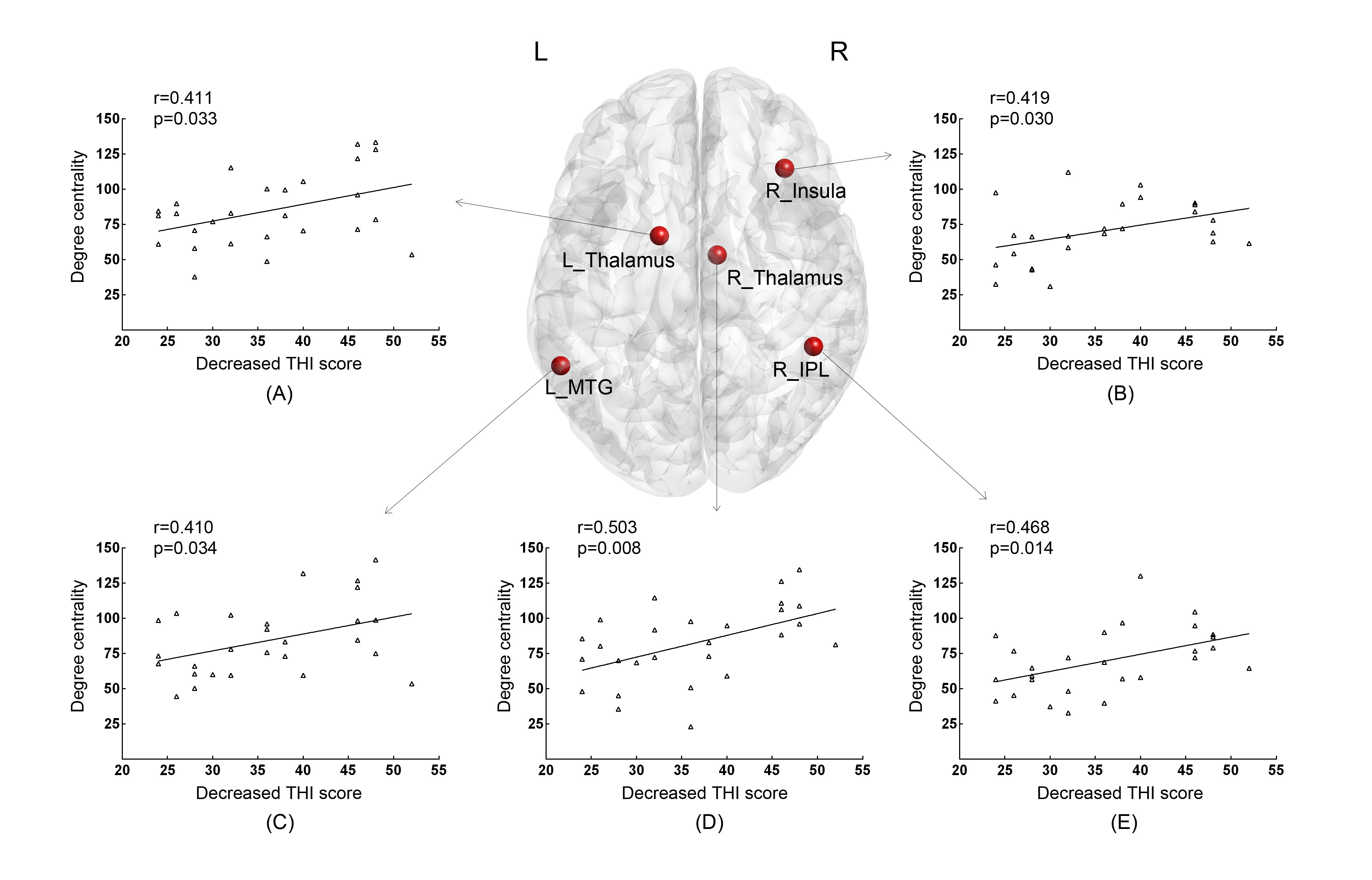
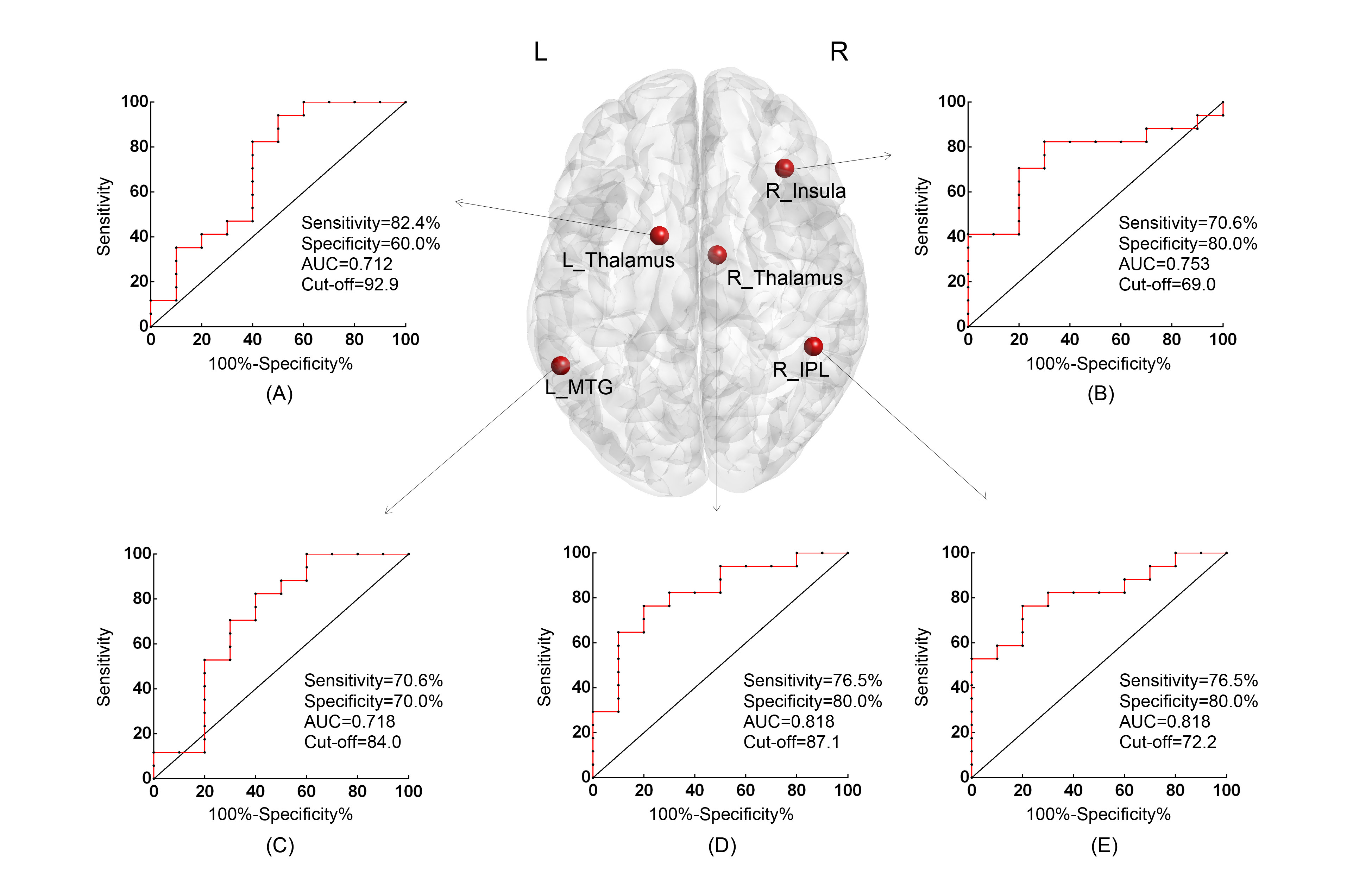
The DC value of ten brain nodes in tinnitus patients were significant increased at baseline. Five nodes, including right insula, inferior parietal lobule (IPL), bilateral thalamus and left middle temporal gyrus, exhibited significant correlations of their DC at baseline with treatment-induced THI changes in tinnitus patients. Receiver operating characteristic curve analyses revealed well performance of the five brain regions in classifying better effect of therapy. Area under the curve (AUC) of the right IPL and thalamus reached the highest value (AUC=0.818), with identical sensitivity and specificity of 80.0% and 76.5%. Moreover, the right thalamus was identified as the optimal regressor as determined by stepwise regression.Conclusion
Our study further supported the involvement of fronto-parietal-cingulate network in mediating tinnitus, and provided DC value of the right thalamus at baseline as an object neuroimaging-based indicator for predicting better efficacy of masking therapy.Acknowledgements
No acknowledgement found.References
1. Bauer, C.A., Tinnitus. New England Journal of Medicine, 2018. 378(13): p. 1224-1231.
2. Carpenter-Thompson, J.R., S.A. Schmidt and F.T. Husain, Neural Plasticity of Mild Tinnitus: An fMRI Investigation Comparing Those Recently Diagnosed with Tinnitus to Those That Had Tinnitus for a Long Period of Time. Neural Plast, 2015. 2015: p. 161478.
3. Lv, H., et al., Resting-State Functional MRI: Everything That Nonexperts Have Always Wanted to Know. AJNR Am J Neuroradiol, 2018. 39(8): p. 1390-1399.
4. Ueyama, T., et al., Alterations of Regional Cerebral Blood Flow in Tinnitus Patients as Assessed Using Single-Photon Emission Computed Tomography. Plos One, 2015. 10(9): p. e0137291.
5. De Ridder, D., Vanneste, S., et al., An integrative model of auditory phantom perception: tinnitus as a unified percept of interacting separable subnetworks. Neurosci Biobehav Rev, 2014. 44: p. 16-32.
Figures