3890
Fractional amplitude of low frequency fluctuation associated with chess expertise: possible role of insular cortex1101 Nicolls Road, Stony Brook Medicine, Stony Brook, NY, United States, 2Biomedical engineering, Stony Brook University, Stony Brook, NY, United States, 3Radiology, Stony Brook Medicine, Stony Brook, NY, United States
Synopsis
While various expertise domains have been studied extensively, the literature body provides only a few reports on chess expertise at rest. To fill the literature gap of resting-state study on chess expertise, we fALFF differences on a dataset consists of 29 chess experts and 29 novices and found a higher fALFF in the right
Introduction
While task-based and resting-state fMRI (rsfMRI) has been used to study many expertise domains (such as radiology, gymnastics, sports, and musics), only a few studies investigated chess expertise domain. Differences in caudate volume [1,2], gray matter volume and cortical thickness in the occitpio-temporal junction and precunei [3] have been reported between chess experts and novices. Task-based fMRI reported differences in activation in the bilateral collateral sulci and bilateral retrosplenial cortices when viewing distorted chessboards [4], and higher activation in the temporo-parietal junction associated with better gestalt perception of chessboards [5]. To our knowledge, there was a single report of resting-state fMRI study on chess expertise [1]. In that study, the authors used caudate as a seed and found altered functional connectivity (FC) to the default mode network. In this study, we employed fractional amplitude of low-frequency fluctuation (fALFF) and seed-to-voxel functional connectivity analysis to investigate whether there are functional network differences between chess experts and novices. fALFF was chosen over ALFF because it has been suggested that fALFF is more sensitive to local difference [6]. Seed-to-voxel functional connectivity analysis was performed using fALFF results as seeds.Method
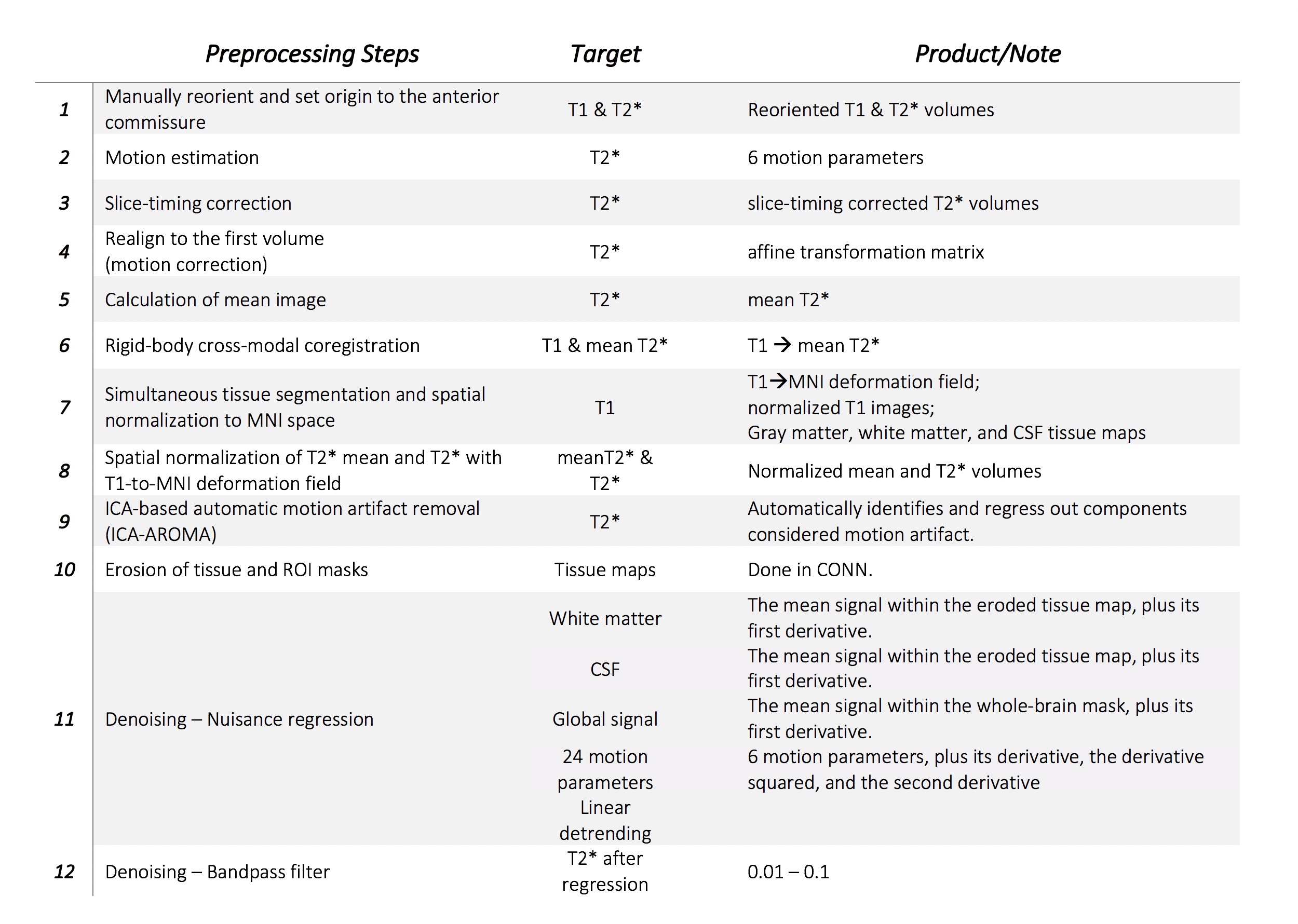
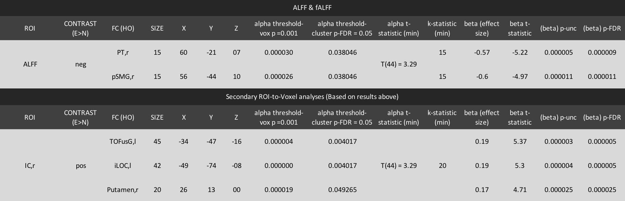
Study was performed on 29 professional Chinese chess masters and grand-masters (ELO rating >2200) and 29 novices who played chess at a regular basis. T1-weighted MRI (1mm isotropic, FOV=176x256x256mm) and EPI (TR=2 sec, 3.75x3.75x5mm, FOV=240x240x150mm, 205 volumes) were obtained from an open dataset [7]. All participants were right-handed, with no history of neurological disorders upon data collection. Data were preprocessed using SPM12. EPI volumes of each participants were corrected for slice-timing and motion, coregistered within subjects, segmented for tissue masks, normalized to MNI space, and smoothed with 8 mm gaussian kernel. Denoising, nuisance regression and statistical analysis were conducted with an SPM toolbox CONN [8]. Detailed preprocessing steps please refer to Table1. Seven experts and five novices were removed from analysis due to excessive motion artifact, corrupted T1-weighted volumes, or unknown cause of different TR and slice numbers of T2* volumes. We first investigated the group difference in fALFF [9] between experts and novices, then further conducted a secondary seed-to-voxel correlational analysis based on a significant result found in fALFF analyses. All the results were first thresholded at p=0.001 at each voxel, the surviving voxels were further thresholded with a cluster threshold at FDR-corrected p=0.05. Seed definition and anatomical reference were based on the Harvard-Oxford atlas in FSL.Results
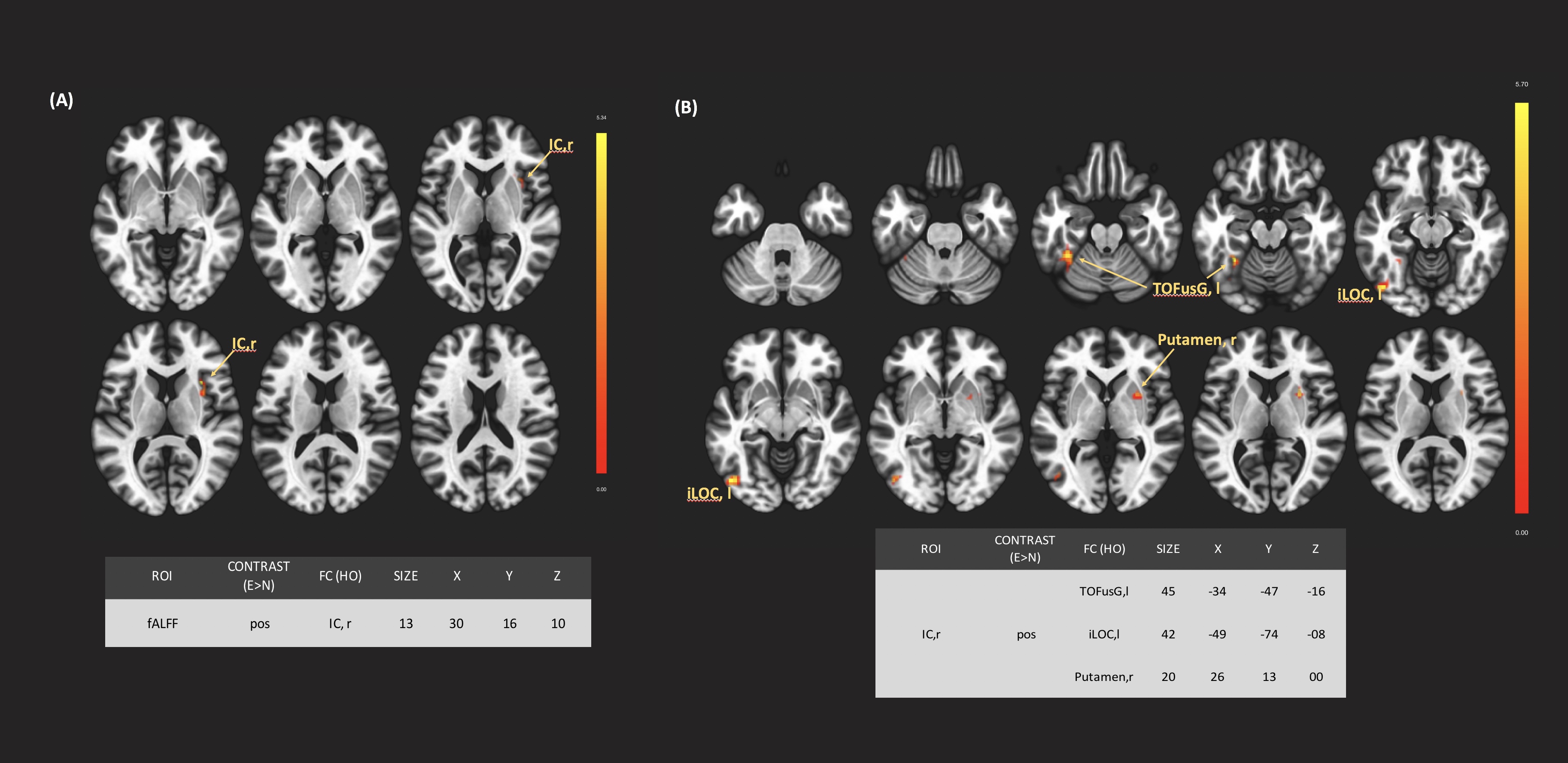
Experts showed higher fALFF in the right insula (IC,r) compared to novices (Figure 1). With IC,r as a seed, experts showed higher FC to the left temporo-occipital fusiform gyrus (TOFusG,l), the left inferior lateral occipital cortex (iLOC,l), and the right putamen (Figure 1). Statistics and finds are summarized in Table 2.Discusion
The insula is a key structure in the salience network and has been implicated in redirecting attention to meaningful stimuli. The ability to flexibly attend to and switch between behaviorally salient stimuli is crucial for generating appropriate behavioral responses, such as recognizing key chess pieces to generate the next best move in a chess game. A higher fALFF found in this region suggest its involvement in chess expertise. The iLOC and the TOFusG is well-known to accommodate regions involving object recognition of faces, scenes, body parts, and objects of expertise [10,11,12,13]. The increased connectivity from insula to these two structures suggest its involvement in chess expertise. The putamen is a part of the dorsal striatum consists of heterogenous compartments, playing various roles in motor control [14], learning [15], and executive functions [16]. It integrates information via the cortico-striato-cortical loop. The putamen might serve as a communicator between the insula and the two regions involving in visual processing: TOFusG and iLOC.Conclusion
Chess experts showed increased fALFF in the insula, and higher functional connectivity from the insula to the left inferior lateral occipital cortex and the right putamen, which are involved in visual recognition. These findings suggest that an enhanced functional integration amongst these regions may be important expert performance. We speculate that stronger connectivity of the hub of salience network and visual processing areas enables experts to exhibit high performance.Acknowledgements
NoneReferences
1. Duan, Xujun, Wei Liao, Dongmei Liang, Lihua Qiu, Qing Gao, Chengyi Liu, Qiyong Gong, and Huafu Chen. “Large-Scale Brain Networks in Board Game Experts: Insights from a Domain-Related Task and Task-Free Resting State.” PLOS ONE 7, no. 3 (March 12, 2012a): e32532. https://doi.org/10.1371/journal.pone.0032532.
2. Duan, Xujun, Sheng He, Wei Liao, Dongmei Liang, Lihua Qiu, Luqing Wei, Yuan Li, Chengyi Liu, Qiyong Gong, and Huafu Chen. “Reduced Caudate Volume and Enhanced Striatal-DMN Integration in Chess Experts.” NeuroImage 60, no. 2 (April 2, 2012b): 1280–86. https://doi.org/10.1016/j.neuroimage.2012.01.047.
3. Hänggi, Jürgen, Karin Brütsch, Adrian M. Siegel, and Lutz Jäncke. “The Architecture of the Chess Player’s Brain.” Neuropsychologia 62 (September 2014): 152–62. https://doi.org/10.1016/j.neuropsychologia.2014.07.019.
4. Bartlett, James, Amy L. Boggan, and Daniel C. Krawczyk. “Expertise and Processing Distorted Structure in Chess.” Frontiers in Human Neuroscience 7 (2013). https://doi.org/10.3389/fnhum.2013.00825. 5. Rennig, Johannes, Merim Bilalic, Elisabeth Huberle, Hans-Otto Karnath, and Marc Himmelbach. “The Temporo-Parietal Junction Contributes to Global Gestalt Perception—Evidence from Studies in Chess Experts.” Frontiers in Human Neuroscience 7 (2013). https://doi.org/10.3389/fnhum.2013.00513.
6. Zou, Qi-Hong, Chao-Zhe Zhu, Yihong Yang, Xi-Nian Zuo, Xiang-Yu Long, Qing-Jiu Cao, Yu-Feng Wang, and Yu-Feng Zang. “An Improved Approach to Detection of Amplitude of Low-Frequency Fluctuation (ALFF) for Resting-State FMRI: Fractional ALFF.” Journal of Neuroscience Methods 172, no. 1 (July 15, 2008): 137–41. https://doi.org/10.1016/j.jneumeth.2008.04.012.
7. Li, Kaiming, Jing Jiang, Lihua Qiu, Xun Yang, Xiaoqi Huang, Su Lui, and Qiyong Gong. “A Multimodal MRI Dataset of Professional Chess Players.” Scientific Data 2 (September 1, 2015). https://doi.org/10.1038/sdata.2015.44.
8. Whitfield-Gabrieli, Susan, and Alfonso Nieto-Castanon. “Conn: A Functional Connectivity Toolbox for Correlated and Anticorrelated Brain Networks.” Brain Connectivity 2, no. 3 (May 29, 2012): 125–41. https://doi.org/10.1089/brain.2012.0073.
9. Cole, David M., Stephen M. Smith, and Christian F. Beckmann. “Advances and Pitfalls in the Analysis and Interpretation of Resting-State FMRI Data.” Frontiers in Systems Neuroscience 4 (2010). https://doi.org/10.3389/fnsys.2010.00008.
10. Grill-Spector, Kalanit, Zoe Kourtzi, and Nancy Kanwisher. “The Lateral Occipital Complex and Its Role in Object Recognition.” Vision Research 41, no. 10 (May 1, 2001): 1409–22. https://doi.org/10.1016/S0042-6989(01)00073-6.
11. Haushofer, Johannes, Margaret S. Livingstone, and Nancy Kanwisher. “Multivariate Patterns in Object-Selective Cortex Dissociate Perceptual and Physical Shape Similarity.” PLOS Biology 6, no. 7 (July 29, 2008): e187. https://doi.org/10.1371/journal.pbio.0060187.
12. Eger, Evelyn, John Ashburner, John-Dylan Haynes, Raymond J. Dolan, and Geraint Rees. “FMRI Activity Patterns in Human LOC Carry Information about Object Exemplars within Category.” Journal of Cognitive Neuroscience 20, no. 2 (October 25, 2007): 356–70. https://doi.org/10.1162/jocn.2008.20019.
13. Lerner, Y., T. Hendler, and R. Malach. “Object-Completion Effects in the Human Lateral Occipital Complex.” Cerebral Cortex 12, no. 2 (February 1, 2002): 163–77. https://doi.org/10.1093/cercor/12.2.163.
14. Wymbs, Nicholas F., Danielle S. Bassett, Peter J. Mucha, Mason A. Porter, and Scott T. Grafton. “Differential Recruitment of the Sensorimotor Putamen and Frontoparietal Cortex during Motor Chunking in Humans.” Neuron 74, no. 5 (June 7, 2012): 936–46. https://doi.org/10.1016/j.neuron.2012.03.038.
15. Lovinger, David M. “Neurotransmitter Roles in Synaptic Modulation, Plasticity and Learning in the Dorsal Striatum.” Neuropharmacology 58, no. 7 (June 1, 2010): 951–61. https://doi.org/10.1016/j.neuropharm.2010.01.008.
16. Beilen, Marije van, and Klaus L. Leenders. “Putamen FDOPA Uptake and Its Relationship Tot Cognitive Functioning in PD.” Journal of the Neurological Sciences, Dementia in Parkinson’s Disease: International Symposium, 248, no. 1 (October 25, 2006): 68–71. https://doi.org/10.1016/j.jns.2006.05.033.
Figures