3883
Hemisphere-specificity between global synchronization of spontaneous brain activity and movement function in Hippocampus of Parkinson’s disease patients: a resting-state fMRI study1Institute of Biomedical and Health Engineering, Shenzhen Institutes of Advanced Technology, Chinese Academy of Sciences, Shenzhen, China, 2Deep Bay Innovation Co., Ltd., Shenzhen, China, 3Department of Neurology, Guangzhou First People’s Hospital, Guangzhou, China, 4Department of Radiology, Guangzhou First People’s Hospital, Guangzhou, China
Synopsis
This study enrolled age-match of 36 people with PD and 35 normal controls (NC) in resting-state functional MRI. Altered global signal synchronizations in PD were explored, and correlations between global synchronizations and clinical assessments (cognitive and movement functions) were investigated. Alterations of global synchronizations were observed in left postcentral gyrus (PostC), left supramarginal gyrus and right opercular-inferior frontal gyrus in people with PD. The correlation results imply postcentral gyrus and hippocampus as critical brain regions associated with both cognitive performances and movement functions in PD.
INTRODUCTION
Abnormalities of cognitive performances and movement functions are widely reported in people with Parkinson’s disease (PD) 1. The mechanisms therein are complicated and assumed to be associated with a coordination of various brain regions. This study explored the altered global synchronizations of spontaneous brain activities and investigated the neural correlations of cognitive performances and movement function in people with PD.
METHODS
Age-match of 36 people with PD and 35 normal controls (NC) were enrolled in resting-state functional MRI scanning, lying quietly and stayed awake with eye closed. Functional image and structural T1 image were both obtained from all subjects. Data preprocessed were performed in DPABI that implemented in MATLAB. Degree of centrality (DC) was calculated for both two groups to measure the global synchronization of brain activity 2. To evaluate group differences, two sample t-test was performed on the DC maps of NC and PD, within gray matter mask, age and head motion of FD Jenkinson 3 as covariates, permutation test with threshold-free cluster enhancement which reaches the best balance between family-wise error rate ( under 0.05) and test-retest reliability 4. The value of DC within the brain regions showing significant group DC differences were extracted, and then correlated with the clinical assessments respectively, including: disease duration, Hoehn-Yahr (HY) which measures the severity of PD, cognitive functions measured by frontal assessment battery (FAB) and Montreal cognitive assessment (MOCA), and movement function scores (Unified Parkinson’s Disease Rating Scale, UPDRS-III). The UPDRS-III includes the movement functions of the left/right/middle/overall body. Additionally, DC signals were also extracted form Anatomical Automatic Labeling (AAL116) templates and then correlated with the above clinical assessments to investigate the neural correlations.RESULTS
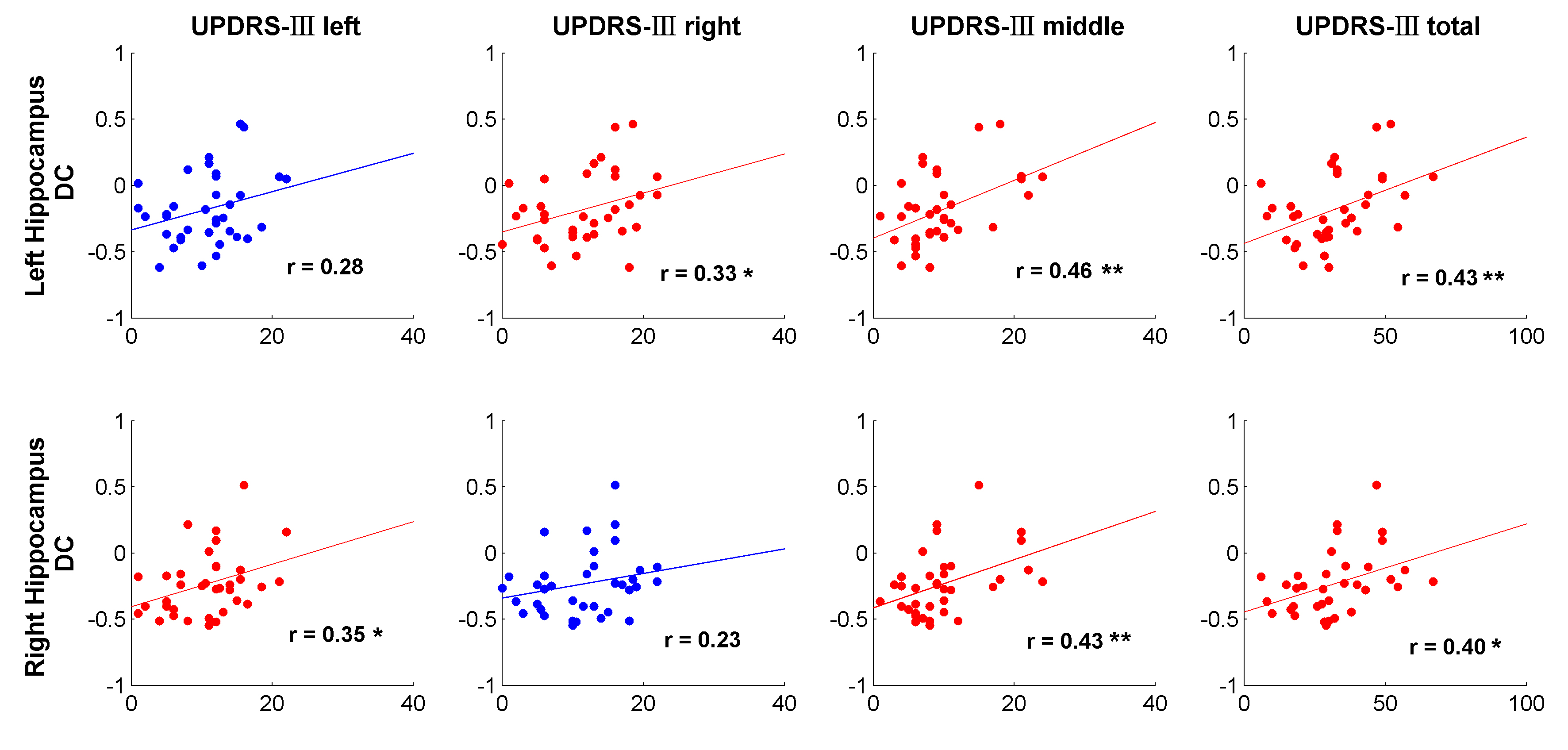
Compare with NC, PD group were observed with decreased DC in left postcentral gyrus (PostC), left supramarginal gyrus and right opercular-inferior frontal gyrus (Figure 1A). PD were observed with significantly (p<0.0001) decreased FAB and MOCA (Figure 1B). Within the significant regions, both FAB and MOCA were demonstrated positive correlations with DC in NC in left PostC, while the results were insignificant in PD (Figure 1C,1D). Correlation analysis of AAL templates showed negative correlations between FAB of NC and DC in hippocampus, while no correlations in PD (Figure 2). In PD, positive correlations were observed between DC of bilateral hippocampus and disease duration, HY (Figure 2) as well as overall movement functions (Figure 3). Interestingly, DC in left hippocampus was positively correlated with UPDRS-III right, and right hippocampus of DC was positivity correlated with UPDRS-III left (Figure 3). While there were no hemispheric bias in correlation with UPDRS-III middle and UPDRS-III total (Figure 3).DISCUSSION
People with PD are characterized with movement disorder. PostC is a conventionally sensorimotor area associated with movement functions. Decreased global synchronizations in left PostC revealed dysfunctional movement in PD. Global signal synchronizations in motor areas (PostC) were observed negatively correlated with cognitive functions (FAB, MOCA) in NC, however, decoupled results were shown in PD (Figure 1C,1D). These findings suggested there exists underlying interactions between cognitive functional and movement functional in PD. Correlation results between DC in hippocampus and various clinical assessments in PD (Figure 2, Figure3) indicated that hippocampus is a critical area in both cognitive and movement performances of PD. Moreover, brain activities of global synchronizations in hippocampus may be associated with hemispheric-specificity movement functions.CONCLUSION
This study provides new insights on the interaction among global coordination of brain activity, cognitive function and movement function in PD. It implies postcentral gyrus and hippocampus as critical brain regions associated with both cognitive performances and movement functions in PD.Acknowledgements
This work is supported in part by grants from the National Key Research and Develop Program of China (2016YFC0105102), the Leading Talent of Special Support Project in Guangdong (2016TX03R139), the Shenzhen Key Technical Research Project (JSGG20160229203812944), the Science Foundation of Guangdong (2017B020229002, 2015B020233011, 2014A030312006) and the Beijing Center for Mathematics and Information Interdisciplinary Sciences, and the National Natural Science Foundation of China (61871374).References
1.Kudlicka A, Clare L, Hindle J V. Executive functions in Parkinson’s disease: Systematic review and meta-analysis. Mov Disord. 2011;26(13):2305-2315. doi:10.1002/mds.23868
2.Buckner RL, Sepulcre J, Talukdar T, et al. Cortical Hubs Revealed by Intrinsic Functional Connectivity: Mapping, Assessment of Stability, and Relation to Alzheimer’s Disease. J Neurosci. 2009;29(6):1860-1873. doi:10.1523/JNEUROSCI.5062-08.2009
3.Jenkinson M, Bannister P, Brady M, Smith S. Improved optimization for the robust and accurate linear registration and motion correction of brain images. Neuroimage. 2002;17(2):825-841. doi:10.1016/S1053-8119(02)91132-8
4. Chen X, Lu B, Yan C. Reproducibility of R-fMRI Metrics on the Impact of Different Strategies for Multiple Comparison Correction and Sample Sizes. Hum Brain Mapp. 2018;39(July 2017):300-318. doi:10.1002/hbm.23843
Figures