3882
Weaker Intra- and Inter-nodal Connectivity of Resting State Networks in Drug-naive Attention-Deficit/Hyperactivity Disorder1Department of Radiology, Huaxi Magnetic Resonance Research Centre (HMRRC), Chengdu, China, 2Department of Psychiatry, The First Affiliated Hospital of Wenzhou Medical University, Wenzhou, China
Synopsis
We used group-level independent component analysis (ICA) to investigate functional network connectivity in drug-naïve ADHD children. We extracted eight resting state networks and found that lower intranodal and internodal functional connectivity among executive control, precuneus and default mode networks in ADHD children compared to controls, especially located in prefrontal and parietal regions. This finding may provide new insights into functional connectivity alterations in ADHD children and promote the exploration of functional network connectivity in future researches.
INTRODUCTION
Attention-deficit/hyperactivity disorder (ADHD) is the most common childhood-onset neurobehavioral disorders with 7.2% worldwide-pooled prevalence in children and adolescents according to a latest meta-analysis 1. In recent years, ADHD are increasingly viewed from impairments among distributed neural networks or circuits, which renders resting state functional connectivity (rsFC) an important role in the elucidation of the psychopathology of this disorder. However, many rsFC studies based on predefined regions and rarely discussed functional network connectivity. Thus, we attempted to evaluate resting state network connectivity using independent components analysis (ICA) 2, a data-driven blind source separation approach that allows a model-free analysis of whole-brain functional MRI data without the requirement of seed region selection.METHODS
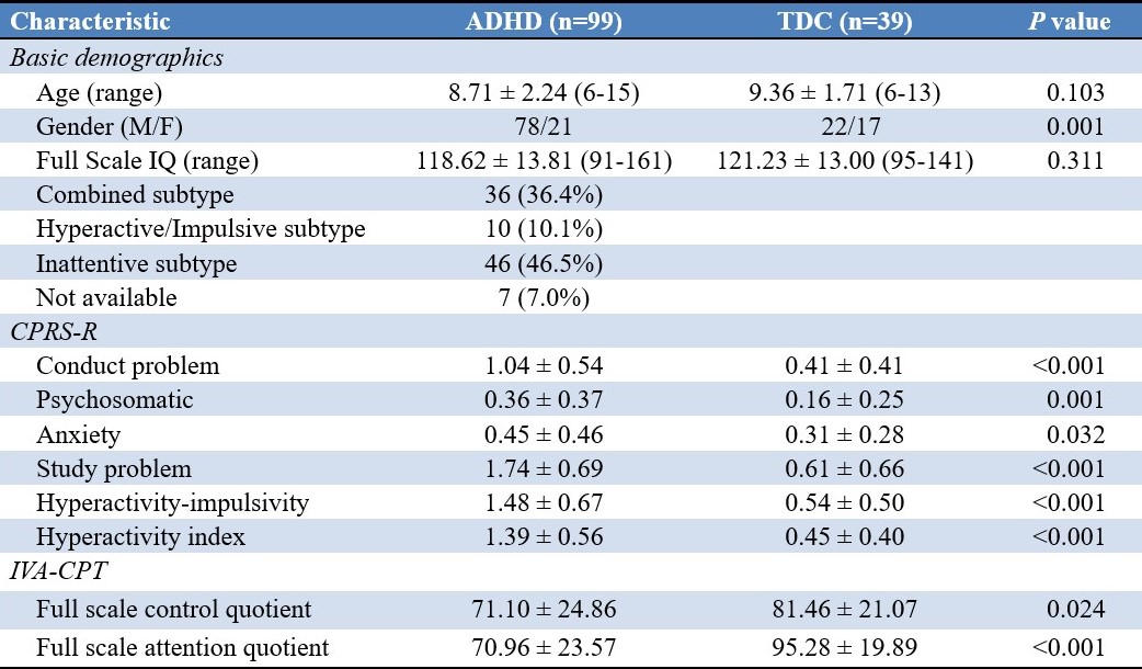
Nighty-nine medication-naive children diagnosed as ADHD according to DSM-V criterion and thirty-nine age and IQ matched typical develop controls (TDC) were recruited. The revised Conners' Parent Rating Scale (CPRS-R) and the Integrated Visual and Auditory Continuous Performance Test (IVA-CPT) were used to assess behavioral performance and attention respectively. This study was approved by the local ethical committee, and written informed consent was obtained from guardians of all subjects.
Resting state functional MR images were obtained on a GE 3T scanner with an eight-channel phased-array head coil, using a gradient-echo echo-planar imaging sequence with slice thickness = 4mm, slice gap = 0.2mm, repetition time = 2000ms, echo time = 30ms, flip angle = 90°, matrix size = 64×64, field of view = 192×192mm2. The fMRI data were preprocessed in SPM8 using an automated pipeline. Image time points with framewise displacement (FD) > 0.2mm was considered severely contaminated with head motion. Functional connectivity was analyzed using a group-level spatial ICA in the GIFT toolbox 3. Data were decomposed into a fixed set of 40 independent components which were subsequently categorized into different intrinsic connectivity networks (ICNs) based on spatial correlation with priori templates 4 and then we visually confirmed the results. Two sample t-tests were applied to compare each selected component spatial maps which were implemented using the statistical capabilities of SPM8 that were accessed through the utilities option in the GIFT software. Functional network connectivity (FNC) correlation matrix was created to investigate functional connectivity between networks using the MANCOVA toolbox. All results were family-wise error (FWE) corrected (P < 0.05).
RESULTS
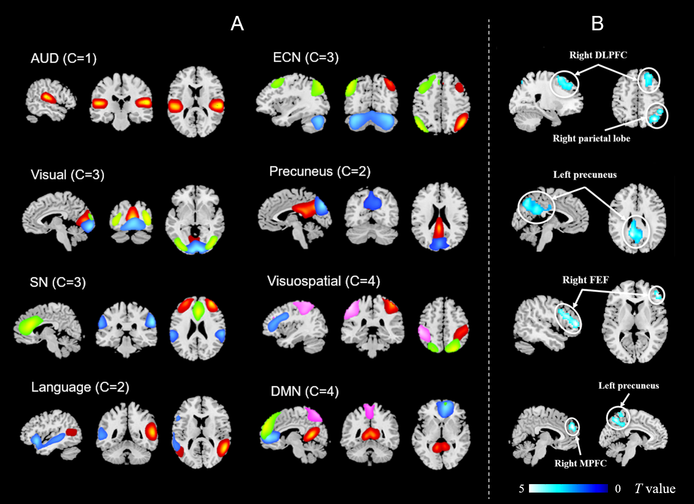
Intranodal Connectivity
We identified eight ICNs with 22 components of ADHD and HC groups. Compared with TDC, ADHD children yielded significant lower intranodal connectivity in clusters located in executive control network (ECN), default mode network (DMN) and visuospatial networks (VSN) (Figure 1, FWE corrected). There were no regions showing higher intranodal connectivity in any of the eight ICNs. In addition, the full scale control quotient of IVA-CPT was negatively correlated with connectivity strength of ECN, DMN and VSN nodes, peaking in the right dorsolateral prefrontal cortex (DLPFC), the left precuneus, the right frontal eye field (FEF) and the right parietal lobe.
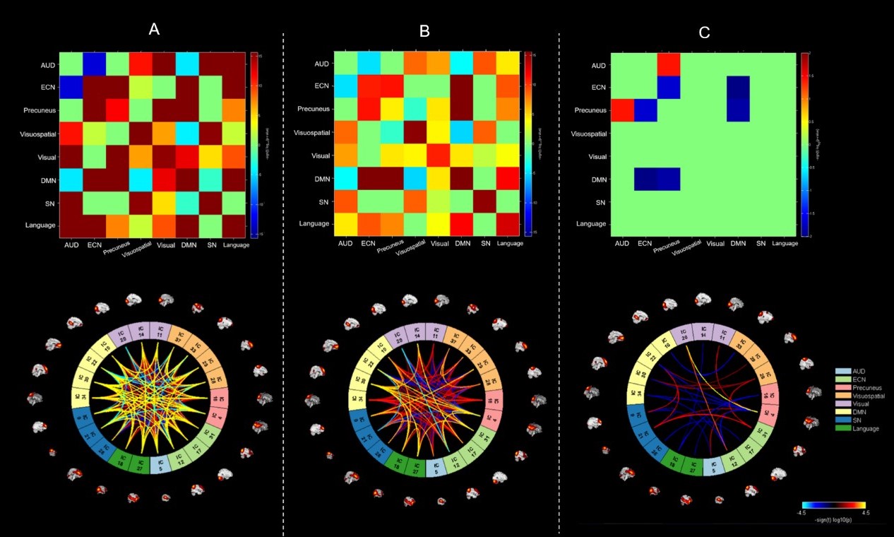
Internodal Connectivity
Comparing to TDC, ADHD children showed weaker internodal connectivity among most of the dominant resting state networks, especially between prefrontal and parietal regions located in the ECN, DMN and precuneus networks (Figure 2).
DISCUSSION&CONCLUSION
In a relatively large group of drug-naive ADHD children, we found both weaker intranodal and internodal rsFC among ECN and DMN, while VSN demonstrates only weaker intranodal connectivity and precuneus network has reduced internodal connectivity with prefrontal and parietal regions. The decreased functional connectivity or connectivity strength within the ECN may cause inhibitory control problems, which lead to symptoms of hyperactivity and impulsivity. However, this result differs from many previous findings that ADHD patients showed increased connectivity within this network 5. As our children had not been exposed to stimulants, we propose this is more likely to reflect the true functional alteration of ADHD children.
Although precuneus has been thought to be the “core node” of the DMN, its involvement in the DMN remains questioned 6. The precuneus and the DMN were identified to be two different ICNs in the present study. The ICA results showed that only the ventral part of the precuneus overlap with the DMN, which is similar to the finding of a previous study 7. Lower intranodal connectivity in precuneus along with lower connectivity between the precuneus and DMN and ECN reflect aberrant functioning in self-referential thought and attention which maybe contribute to the symptom of ADHD.
Acknowledgements
This study was supported by the National Natural Science Foundation (Grant No. 81671669), Science and Technology Project of Sichuan Province (Grant No. 2017JQ0001).References
1. Thomas R, Sanders S, Doust J et al. Prevalence of attention-deficit/hyperactivity disorder: a systematic review and meta-analysis. Pediatrics. 2015; 135 (4): e994-1001
2. McKeown M, Makeig S, Brown G, et al. Analysis of fMRI data by blind separation into independent spatial components. Hum Brain Mapp. 1998; 6: 160-188.
3. Calhoun V, Adali T, Pearlson G, and Pekar J. Group ICA of Functional MRI Data: Separability, Stationarity, and Inference. Proceedings, ICA, San Diego, CA, 2001.
4. Shirer W, Ryali S, Rykhlevskaia E, et al. Decoding subject-driven cognitive states with whole-brain connectivity patterns. Cereb Cortex. 2012; 22 (1): 158-65
5. Castellanos F & Aoki Y. Intrinsic Functional Connectivity in Attention-Deficit/Hyperactivity Disorder: A Science in Development. Biol Psychiatry Cogn Neurosci Neuroimaging. 2016; 1: 253-261.
6. Margulies D, Vincent J, Kelly C, et al. Precuneus shares intrinsic functional architecture in humans and monkeys. PNAS. 2009; 106 (47): 20069–74.
7. Zhang S & Li C. Functional connectivity mapping of the human precuneus by resting state fMRI. NeuroImage. 2012; 59 (4): 3548–3562.
Figures