3877
Functional Domain Connectivity Strength in SchizophreniaVictor M Vergara1 and Vince D Calhoun1
1The Mind Research Network, Albuquerque, NM, United States
Synopsis
Functional network connectivity (FNC) has been successfully used to detect dysfunctions on the brain of schizophrenia patients. However, this work proposes the idea that effects can be observed when studying functional domains (groups of functionally related brain areas). A set of previously unobserved dysfunctions were detected by studying the domain connectivity strength. Domain connectivity is an important framework for future research of functional connectivity.
Introduction
Functional network connectivity among different areas in the brain revealed the existence of abnormalities in the brain of schizophrenia patients (Nelson, Bassett, Camchong, Bullmore, & Lim, 2017; Ray et al., 2017; Zhuo et al., 2018). Effort has been made in identifying these abnormalities in specific brain networks (Zhuo et al., 2018) or using a whole brain framework (Damaraju et al., 2014). The aim of this work is to use functional network connectivity estimated from resting state fMRI data to study schizophrenia abnormalities related to functional groups of brain areas. A functional related group of brain networks will be designated as a domain. Considering single network level as the finest and whole brain as the coarsest, functional domains are an intermediate spatial level.Methods
Data for this work was obtained from a previously publish study of functional network connectivity (FNC) in schizophrenia (Damaraju et al., 2014). Subject pool consists of 163 healthy controls (HC) and 151 schizophrenia patients (SZ) with similar mean age and gender. Collected fMRI data was preprocessed using a pipeline that included rigid body motion correction, followed by slice-timing correction, despiking, normalization to the Montreal Neurological Institute (MNI) template and smoothing. Group independent component analysis (GICA) was used to estimate an FNC matrix for each subject. GICA components were grouped in the functional domains sub-cortical (SBC), auditory (AUD), visual (VIS), sensorimotor (SEN), a broad set of regions involved in cognitive control and attention (COG), default-mode network (DMN) regions, and cerebellum (CER). The whole brain FNC matrix of each subject was then divided in submatrices following the domain grouping. A domain connectivity strength value was then obtained by averaging all values within the submatrices. A illustration of this procedure can be seen in Figure 1. We finally performed an unpaired t-test looking for significant group differences. We corrected from multiple testing using the false discovery rate (FDR) technique.Results
There were many group differences among the functional domains. The following domain connectivity strengths were lower in schizophrenia: the connectivity among the domains AUD, VIS and SEN; the trio AUD, VIS and SEN (for simplicity let’s rename it as AVS) with the DMN domain; in the subcortical area SCB-SCB, SBC-CER; and finally COG-COG. The following domain connectivity strengths were higher in schizophrenia: the trio AVS with the SBC domain; and AVS with CER. These significant differences can be observed in Figure2 by the red and blue boxes, but the non-significant boxes have been white out.Discussion
This work presents a different way of looking at the functional connectivity. We have incorporated a prior knowledge in resting state to refocus the FNC analysis as brain networks groped on functionally related domains. This new conceptualization has been recently presented in the literature (Miller, Vergara, Keator, & Calhoun, 2016; Vergara, Miller, & Calhoun, 2017), but it is the first time it is applied to connectivity strength in schizophrenia. Domain connectivity strength revealed a large number of effects that were not previously observed in the same data, but with a different spatial focus (Damaraju et al., 2014). Domain analysis found extra abnormalities in schizophrenia: 1) lower within connectivity in subcortical and cognitive domains; 2) lower between connectivity on subcortical-cerebellum and DMN-AVSN; 3) higher between connectivity cerebellum-AVSN. These results are consistent with the previous ones where dysfunctional connectivity between thalamus and AVS was observed (Damaraju et al., 2014). These observations are also consistent with reports in the literature (Woodward, Karbasforoushan, & Heckers, 2012) and it is possible that thalamus was the major contributor to the subcortical results observed in this work. However, other effects have been observed for the first time. Some of these effects may be related with the existence of a disconnection between subcortical regions and cerebellum (Anticevic et al., 2014). Further exploration will be required to explain the other abnormal domain connectivities.Conclusion
Results presented indicate that abnormalities of brain connectivity could be obscured by the spatial level of the analysis. Whole brain analysis might not allow for the discovery of regional dysfunctions. Analysis focusing on small brain regions might be too specific and disregard interactions of pinpointed areas with others. By employing a domain connectivity approach, we have allowed for the analysis of brain areas as a group resulting in the discovery of previously undetected abnormalities in schizophrenia.Acknowledgements
This work was supported by grants from the National Institutes of Health grant numbers 2R01EB005846, R01REB020407, and P20GM103472; and the National Science Foundation (NSF) grants 1539067/1631819 to VDC.References
Anticevic, A., Cole, M. W., Repovs, G., Murray, J. D., Brumbaugh, M. S., Winkler, A. M., . . . Glahn, D. C. (2014). Characterizing thalamo-cortical disturbances in schizophrenia and bipolar illness. Cereb Cortex, 24(12), 3116-3130. doi:10.1093/cercor/bht165 Damaraju, E., Allen, E. A., Belger, A., Ford, J. M., McEwen, S., Mathalon, D. H., . . . Calhoun, V. D. (2014). Dynamic functional connectivity analysis reveals transient states of dysconnectivity in schizophrenia. Neuroimage Clin, 5, 298-308. doi:10.1016/j.nicl.2014.07.003 Miller, R. L., Vergara, V. M., Keator, D. B., & Calhoun, V. D. (2016). A Method for Intertemporal Functional-Domain Connectivity Analysis: Application to Schizophrenia Reveals Distorted Directional Information Flow. IEEE Trans Biomed Eng, 63(12), 2525-2539. doi:10.1109/TBME.2016.2600637 Nelson, B. G., Bassett, D. S., Camchong, J., Bullmore, E. T., & Lim, K. O. (2017). Comparison of large-scale human brain functional and anatomical networks in schizophrenia. Neuroimage Clin, 15, 439-448. doi:10.1016/j.nicl.2017.05.007 Ray, K. L., Lesh, T. A., Howell, A. M., Salo, T. P., Ragland, J. D., MacDonald, A. W., . . . Carter, C. S. (2017). Functional network changes and cognitive control in schizophrenia. Neuroimage Clin, 15, 161-170. doi:10.1016/j.nicl.2017.05.001 Vergara, V. M., Miller, R., & Calhoun, V. (2017). An information theory framework for dynamic functional domain connectivity. J Neurosci Methods, 284, 103-111. doi:10.1016/j.jneumeth.2017.04.009 Woodward, N. D., Karbasforoushan, H., & Heckers, S. (2012). Thalamocortical dysconnectivity in schizophrenia. Am J Psychiatry, 169(10), 1092-1099. doi:10.1176/appi.ajp.2012.12010056 Zhuo, C., Wang, C., Wang, L., Guo, X., Xu, Q., Liu, Y., & Zhu, J. (2018). Altered resting-state functional connectivity of the cerebellum in schizophrenia. Brain Imaging Behav, 12(2), 383-389. doi:10.1007/s11682-017-9704-0Figures
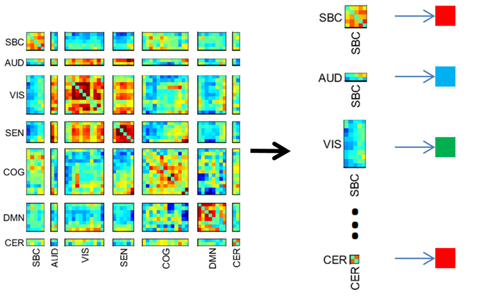
Figure 1
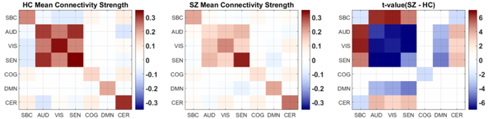
Figure 2