3869
Quantitative Assessment of Temporal Changes in fMRI and DTI Parameters in the Squirrel Monkey Spinal Cord after Injury1Radiology, Vanderbilt University, Nashville, TN, United States
Synopsis
Previous studies have reported changes in resting state functional connectivity between horns in spinal cord following injury. In this work we quantified changes in connectivity and diffusion parameters at identical spinal segments in squirrel monkey cervical spinal cords acquired in concurrent sessions to compare the temporal variations of functional and structural changes during recovery after spinal cord injury (SCI). The degree of axonal disruption and demyelination measured using fractional anisotropy and radial diffusivity at two stages of recovery were compared in various white matter tracts.
Introduction
Correlated fluctuation of low frequency BOLD signals in a resting state between horns of gray matter have been detected in spinal cords in humans, rats and monkeys [1,2,3]. This resting state functional connectivity (rsFC) is weakened following injury but recovers over time [3]. Diffusion tensor imaging (DTI) has been used to measure structural changes along the white matter tracks in the spinal cord and to assess axonal disruption and demyelination [4, 5]. In this work we analyzed the similarity and differences between DTI and rsFC measures along the C4-C8 segments in axial images acquired in the same sessions in squirrel monkeys at 9.4T before and after a targeted unilateral sectioning of the dorsal white matter tract. The temporal changes of DTI parameters of fractional anisotropy (FA) and radial diffusivity (RD) in the injured dorsal track were analyzed and related to changes in rsFC between two pairs of ROIs in grey matter horns, at two time points during recovery after the injury along the cord cross spinal segments.Material and Method
MRI images were acquired with an Agilent 9.4T scanner using a saddle-shaped transmit-receive surface coil of anaesthetized squirrel monkeys. Four animals participated in the study whose dorsal column tract spinal cords were later lesioned on one side at C5 level to acquire post SCI data. A fast gradient echo sequence (flip angle = ~18°, TR = 46.88ms, TE: 6.5ms, 3s per volume) was used to collect resting state functional MRI data (300 dynamics). Spin-echo diffusion sequences were used with an echo planar imaging readout (TR/TE = 3000/33ms, 4 shots, resolution = 0.333x0.333 mm2) with b value = 1000s/mm2(30 directions) for DTI data acquisitions. Identical FOV (32x32mm2) five slices (in 3mm thickness) were selected for both studies. The Fisher transformed z-scores of correlation coefficients of resting state fMRI signal (0.01-0.1Hz) between dorsal-to-dorsal and ventral-to-dorsal gray matter horns were quantified using custom code (AFNI, [1]). The diffusion parameters (FA&RD) at each axial slice were evaluated using a non-linear tensor fitting model (CAMINO). Post lesion data were acquired at two post-injury time points: recovery stage 1 (1-2 weeks) and stage 2 (6-9weeks). Twenty-four resting state functional (2 runs/animal, fMRI) and twelve DTI datasets (1 run/animal) were analyzed.Results
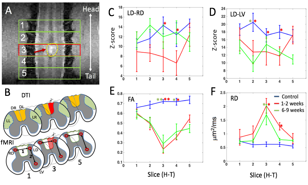
Placements of five axial slice for the data acquisitions are shown in Figure A with a high resolution MTC image obtained from one monkey 2 weeks after a unilateral dorsal column injury in the background. The center slice is positioned around the injury at the left dorsal column (red arrow). A schematic view of the region of interest (ROI) placements in the white (top) and gray matter are shown in Figure B. On each slice, two ROI pairs were selected for rsFC measurements (1. left-right dorsal (LD-RD) and 2. left dorsal-ventral (LD-LV) horns). We analyzed the variations of the FA and RD values of the left dorsal white matter tract over time. Figure C&D show the mean and standard error of z-scores of the correlation coefficients for ROI pairs of 1&2(LD-LR&LD-LV) along the spinal cord segments (slices 1-5) respectively. Figures E&F demonstrate the similar temporal variations of FA and RD on the injured dorsal tract. Statistically significant differences between pre-injury (blue line) recovery stage 1(red line) and stage 2 (green line) were measured using ANOVA1 (** p < 0.005, * < 0.05, indicated by different color stars). Resting state z-scores showed significant differences between pre-injury and recovery stage 1 in comparison to stage 2 in the slice 3 (injury level) and adjacent slices. Additionally, we observed a strong correlation between the mean rsFC z-score (left-right dorsal horn) and the mean RD and AD at the early stage of recovery. The temporal variation of DTI parameters in three other white matter tracts are under investigationDiscussion
This study measured the effects of SCI on axonal integrity of the injured white matter tract and demyelination as well as rsFC between dorsal-dorsal and dorsal-ventral horns and their alterations during the process of recovery. Temporal changes in functional and DTI parameters in combination across spinal segments along the cord are being correlated with behavioral deficits and may be considered as useful predictors and sensitive imaging biomarkers of behavioral recovery.Acknowledgements
This work was supported by a National Institutes of Health grant R01 NS09296 DoD grant W81XWH-17-1-0304 (Gore). We would like to thank Mr. Fuxue Xin and Mrs. Chaoui Tang for their assistance in acquiring the MRI data and animal care.References
1. Barry, R.L., Smith, S.A., Dula, A.N., Gore, J.C., 2014. Resting state functional connectivity in the human spinal cord. eLife 2014;3:e02812. DOI: 10.7554/eLife.02812.
2. Wu, T.L., Wang, F., Mishra Arabinda, Wilson, G. H., Byun, N., Chen, L. M., Gore, J.C., 2018. Resting state functional connectivity in the rat cervical spinal cord at 9.4T. Magn Reson Med. 79(5):2773-83.
3. Chen, L.M., Mishra Arabinda, Yang, P.F., Wang, F., Gore, J.C., 2015. Injury alters intrinsic functional connectivity within the primate spinal cord. Proc Natl Acad Sci U S A 112, 5991-5996.
4. Mishra, Arabinda, Wang, F., Chen, L.M., Gore, J.C., 2017. Spinal DTI parametric changes following traumatic injury in monkeys. 599, May 11-16, Honolulu (USA).
5. Kuhn, F.P., Feydy, A., Launay, N., Colau, M.L., Pairaudeau, S., Laporte, S., Maier, M., A., Lindberg, P., 2016. Kinetic DTI of servical spine diffusivity changes in healthy subjects. Neuroradiology, 58:929-35.
Figures