3864
Olfaction connectivity in Patients with Parkinson’s disease: A comparative fMRI study.1NMR & MRI Facilty, All India Institute of Medical Sciences, New Delhi, India, 2Neurology, All India Institute of Medical Sciences, New Delhi, India, 3All India Institute of Medical Sciences, New Delhi, India, 4Biostatistics, All India Institute of Medical Sciences, New Delhi, India
Synopsis
Olfactory loss is one of the most common non-motor symptoms in Patients with Parkinson’s disease. ROI based functional connectivity analysis was performed on 10 healthy subjects (HC, 6M/4F, mean age=57±4.5 years) and 10 subjects with Parkinson’s disease (9M/1F, mean age=57.7±5.6 years) with olfaction fMRI (floral smell). Altered olfaction linked functional connectivity was present for amygdala, insula and cingulate gyrus in PD. Functional connectivity for olfactory cognition (hippocampus/orbitofrontal) was found intact in both the groups. Primary olfactory structures are more affected compared to the upstream structures, leading to the observed olfactory impairment.
Introduction
Olfactory loss is one of the most common non-motor symptoms in Patients with Parkinson’s disease (PwPD), occurring often in early stages, before the onset of motor symptoms1. Altered processes in the olfactory bulb and olfactory structures within the mesolimbic brain areas are often associated with the ailment2. However, the pattern of olfaction linked activation among PD population is largely unknown. Present study tries to assess connectivity changes in PD compared to healthy individuals using olfaction fMRI task.Methodology
Ten patients with PD (9M/1F, mean age=57.7±5.6 years) and ten healthy controls (HC, 6M/4F, mean age=57±4.5 years) underwent olfaction fMRI with task presented in block design with 15s active smell presentation and 20s rest with airflow. Subjects were scanned in supine position using 3T MR scanner (Achieva, Philips Medical Systems, The Netherlands) with 32 channel head coil. EPI sequence (TR/TE=2000/30ms, slices=40; slice thickness=3.6 mm, gap=0, 310 dynamics) was used to observe the BOLD effects and TFE sequence (TR/TE=8.1/3.7ms, slices=180, slice thickness=1mm) for 3D T1 anatomical imaging. SPM 12 connectivity tool “CONN” (version 15.d) was used for ‘ROI’ based functional connectivity (Fc) analysis. The findings corresponding to the “sweet smell/floral odor” are described here in this article.Results
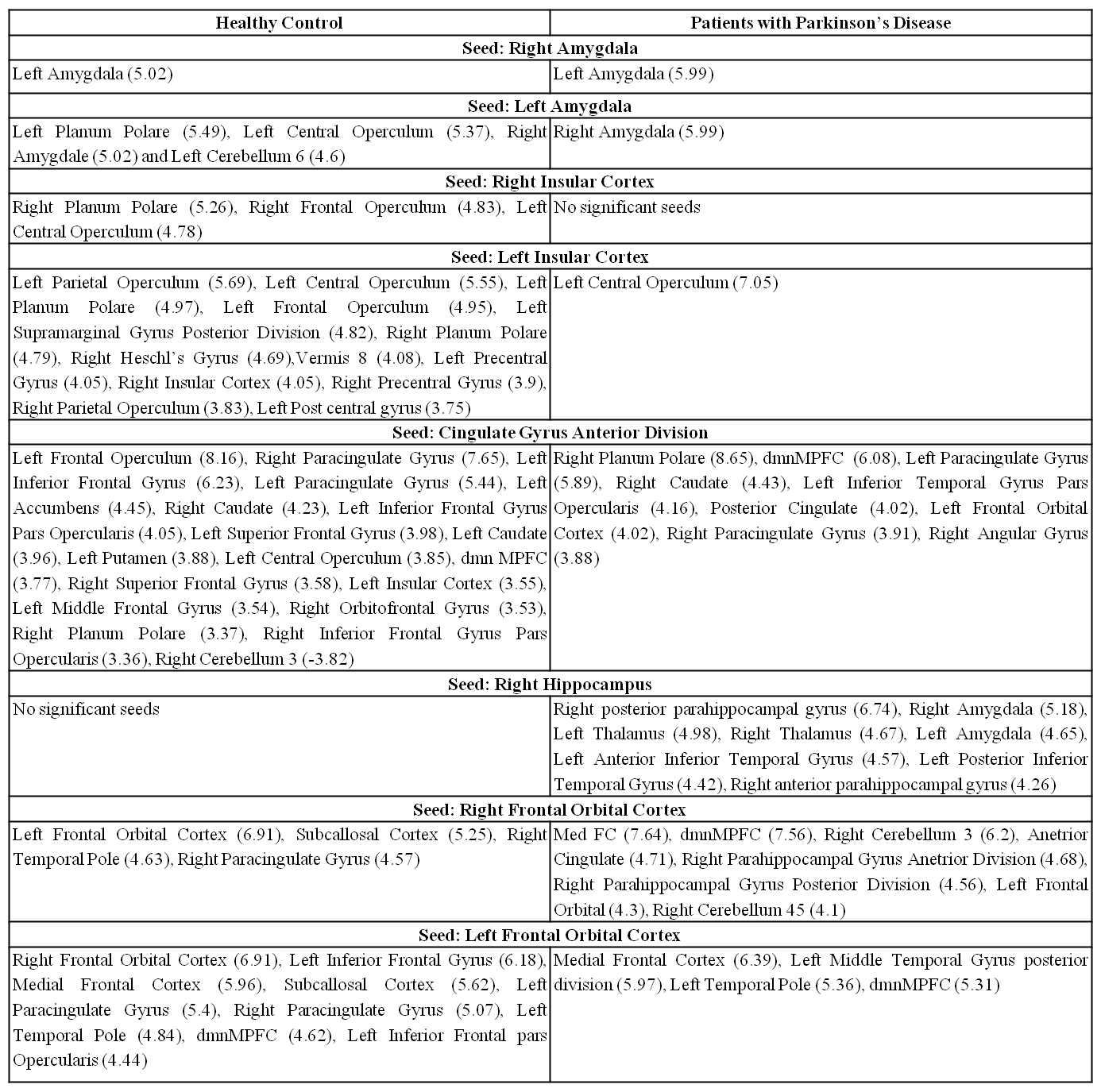
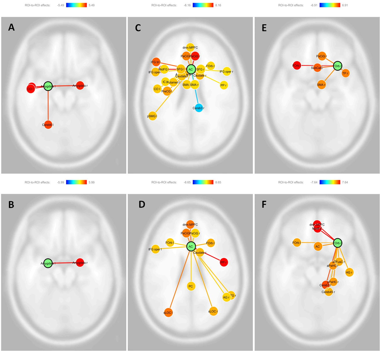
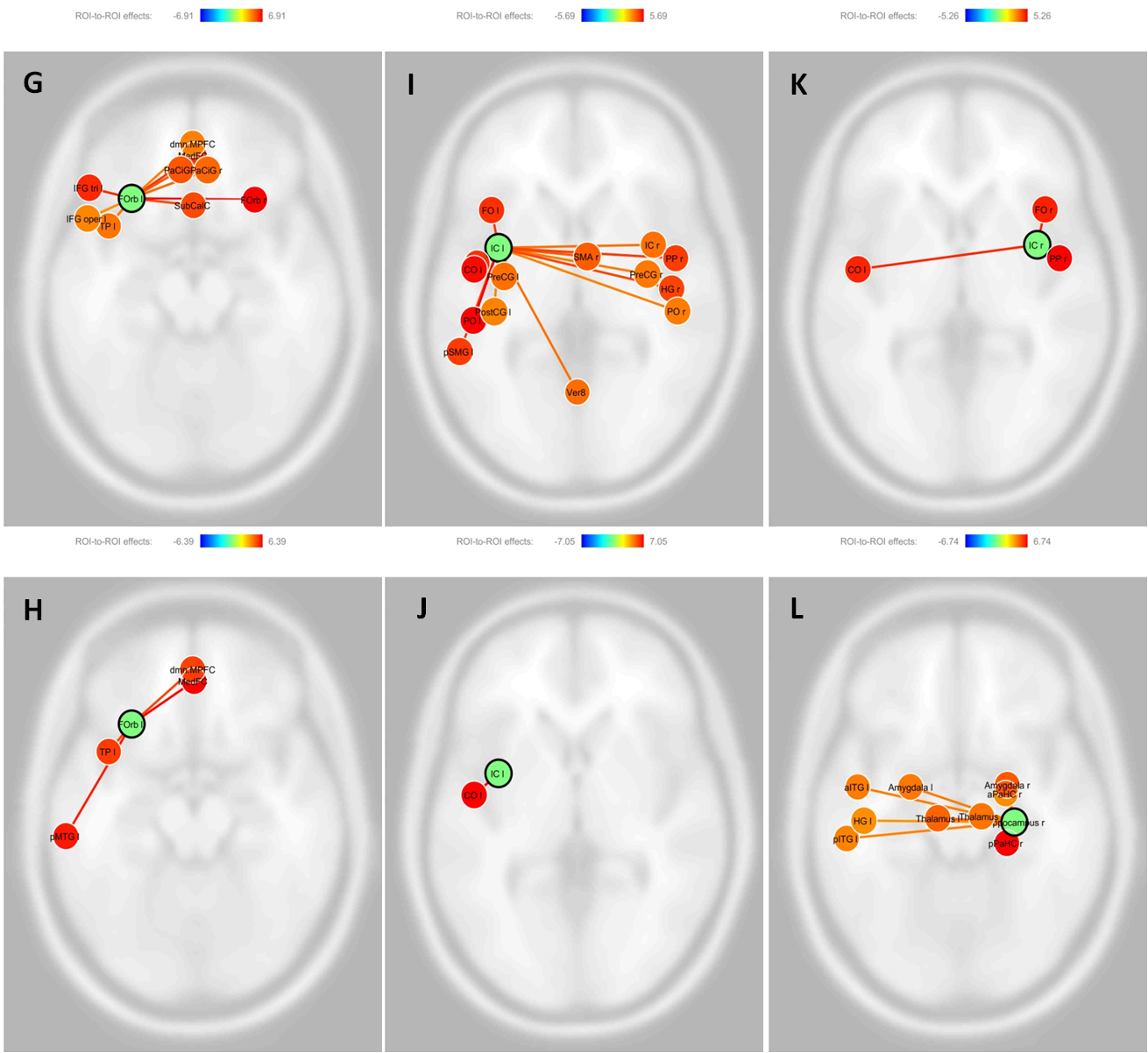
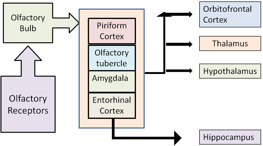
For task based Fc analysis, the seeds were: bilateral amygdala, bilateral insular cortex, cingulate gyrus anterior division, bilateral hippocampus, bilateral frontal orbital cortex (Figures 1,2) as the regions are part of human olfactory circuit3 (Figure 3). For statistical analysis, the threshold was at p-value <0.05 (FDR-corrected) to observe main effects of the two groups. At this level of significance, between group comparison did not give any significant results. Hence, group main effects were compared for individual seeds (Table 1 with the corresponding T-values). 70% of HC and 30% of PD responded correctly for smell identification.Discussion
In patients with PD, amygdala with inter-hemispheric amygdala connectivity only was observed, while HC exhibited connectivity correlation with temporal and cerebellar regions of olfactory roles. Activation of amygdala may be associated with odor intensity and associated emotional response4, which may be impaired in PD. Missing functional connectivity for insular cortex (involved in the processing of gustatory and olfactory inputs5) in PD further adds to this impairment.
Olfactory cognition serves to evaluate general cognition and emerging literature finds correlation for olfaction to cognitive impairment in neurodegenerative conditions6. Considering cognition, anterior cingulate functional connectivity was almost similar for both the groups. However, PD did not reveal connectivity for accumbens, putamen and insula. The orbitofrontal connections, along with other frontal connections, were present in both the groups, suggesting intact olfactory cognition.
The task presented during scanning necessitates identification of the smell (presented). Hippocampus plays a major role in olfaction linked memory formation7 and the region did not exhibit functional connectivity in HC (for both the hemispheres), while only for the right hemisphere in PD. Considering, memory responses from both the groups, this contradicting finding may suggest shorter reaction time in HC leading to habituation or more of frontal and cingulate regions compensating the memory perception in HC.
The seeds amygdala, insula and cingulate gyrus in PwPD miss the major olfaction linked connectivity regions. However, for hippocampus, HC misses most of the functional connections. And for orbitofrontal region, the connectivity was comparable among the two groups, suggesting intactness of olfaction linked cognition for the presented smell in both the groups.
Conclusion
From the findings, we observe that not all the components of central olfactory system are affected in PD during olfaction. The functional connectivity of the primary olfactory structures is affected more compared to the upstream structures, leading to the observed olfactory impairment.Acknowledgements
Funding from SERB (for fellowship of SC) and DST (for the project), Govt. India is acknowledged.References
1. Richardson JT, Zucco GM. Cognition and olfaction: a review. Psychol Bull. 1989; 105: 352-60.
2. Hummel T et al. Olfactory FMRI in patients with Parkinson's disease. Front Integr Neurosci. 2010; 4:125.
3. Purves D, Augustine GJ, Fitzpatrick D, et al., editors. Neuroscience. 2nd edition. Sunderland (MA): Sinauer Associates; 2001.
4. Soudry Y et al. Olfactory system and emotion: common substrates. Eur Ann Otorhinolaryngol Head Neck Dis. 2011; 1:18-23.
5. Veldhuizen MG et al. Identification of human gustatory cortex by activation likelihood estimation. Hum Brain Mapp. 2011; 32:2256-66.
6. Georgiopoulos C et al. Olfactory Impairment in Parkinson's Disease Studied with Diffusion Tensor and Magnetization Transfer Imaging. J Parkinsons Dis. 2017; 7:301-311.
7. Deshmukh SS, Bhalla US. Representation of odor habituation and timing in the hippocampus. J Neurosci. 2003; 23:1903-15.
Figures