3863
Altered Effective Connectivity Anchored in the Posterior Cingulate Cortex and the Medial Prefrontal Cortex in Cognitively Intact Elderly APOE ε4 Carriers: A Preliminary Study1The 2nd Affiliated Hospital of Zhejiang University School of Medicine, Hangzhou, China, 2GE Healthcare Shanghai, Shanghai, China
Synopsis
The APOE ε4 allele is the most reliable genetic risk factor for developing sporadic AD. Based on Granger causality analysis (GCA), we analyzed the APOE ε4-related effects on whole-brain directional connectivity in healthy elderly populations, by setting connected node located in anterior and posterior default mode network (DMN). We found that APOE ε4 carriers had reduced effective connectivity (EC) from the PCC to the anterior and posterior DMN subsystems; additionally, the carriers had increased EC from the parietal cortex to the anterior DMN subsystem. Second, the correlation analyses indicated the decreased EC in the carriers might result from neurofibrillary changes.
Synopsis
The APOE ε4 allele is the most reliable genetic risk factor for developing sporadic AD. Based on Granger causality analysis (GCA), we analyzed the APOE ε4-related effects on whole-brain directional connectivity in healthy elderly populations, by setting connected node located in anterior and posterior default mode network (DMN). We found that APOE ε4 carriers had reduced effective connectivity (EC) from the PCC to the anterior and posterior DMN subsystems; additionally, the carriers had increased EC from the parietal cortex to the anterior DMN subsystem. Second, the correlation analyses indicated the decreased EC in the carriers might result from neurofibrillary changes.Introduction
The APOE ε4 allele is associated with impaired intrinsic functional connectivity in neural networks, especially in the default mode network (DMN). However, effective connectivity (EC) reflects the direct causal effects of one brain region to another, which has rarely been investigated1. Recently, Granger causality analysis (GCA) proved suitable for the study of directionality in neuronal interactions2.Methods
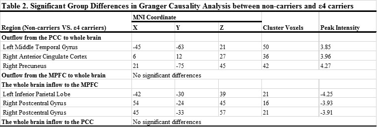
Using GCA, we examined the differences in the EC between the anterior medial prefrontal cortex/posterior cingulate cortex (aMPFC/PCC) and the whole brain in 17 ε4 carrying and 32 non-carrying cognitively intact elderly individuals (voxel-height threshold p<0.01, cluster level p<0.01, Table 1). To be specific, for estimating the time-directed prediction between the time series of a seed region and the rest of the whole brain, a time lag of one TR (3000 ms) was used. Furthermore, correlation analyses were performed between the abnormal EC and cognition/AD-related pathologies (from cerebrospinal fluid, p<0.005).Results
Compared with the non-carriers, the results showed that the ε4 carriers exhibited decreased EC from the PCC to the whole brain in the middle temporal gyrus (MTG), the anterior cingulate cortex (ACC), and the precuneus (PCu). Meanwhile, the ε4 carriers demonstrated increased EC from the whole brain to the aMPFC in the inferior parietal lobe (IPL) and the postcentral gyrus (PCG, Figures 1 and Table 2). Considering the influence of physiologic noise on EC, the results remain mostly unchanged. Furthermore, the correlation analyses suggested that the EC from the IPL/PCG to the aMPFC was related to episodic memory in non-carriers, while the decreased EC from the PCC to the ACC (causal influence from the PCC to the ACC) was associated with increased levels of t-tau (r=-0.80, p<0.001) and p-tau (r=-0.74, p<0.005) in the ε4 carriers (Figure 2).Discussion
As a core hub, the PCC has reciprocal functional connections with each component of the DMN. Previous studies of GCA conducted in healthy volunteers also demonstrated that the PCC works as the primary emitter and terminal receiver of causal influences within the whole brain. The most prominent result of our findings was that there was a reduced causal influence from the PCC to the ACC in ε4 carriers. Supportive neuropathologic evidence comes from the work of Braak et al., who documented that selective distribution of AD-related pathologies begin to aggregate in regions located in the posterior DMN then spread to the front of the brain. Moreover, our results indicated that ε4 allele carriers had reduced causal influence from the PCC to the MTG/PCu. These results suggest that, apart from the disconnection between the anterior and posterior DMN subsystems, ε4 allele carriers still had a regional disconnection within the posterior subsystem of the DMN.
Notably, APOE ε4 carriers had a positive causal influence from the left IPL and the right PCG to the aMPFC, the second hub of the DMN. We interpreted the enhanced driving effects from the IPL/PCG to the aMPFC as the compensation, for the cognitive abilities that should have been disrupted in APOE ε4 carriers. As an exploratory analysis, we further examined the relationship between the mean EC in regions with significant differences between the groups and CSF biomarkers. Our results revealed that the EC of the right ACC (causal influence from PCC to ACC) was strongly related to the levels of t-tau and p-tau in the APOE ε4 carriers. Accordingly, we speculated that deficits in EC might be a consequence of the early events of APOE ε4 allele-mediated reductions in the efficiency of unphosphorylated binding tau.
Conclusion
Our study presented a hypothesized dynamic model of the intrinsic brain network in APOE ε4 carriers; a putative downward system of negative influence could be traced from the PCC to both the posterior and anterior DMN subsystems; meanwhile, the anterior DMN subsystem received positive compensatory influence from the parietal cortex. Finally, we further speculated that APOE ε4 allele-related increases in neurofibrillary tangles might act as an initial factor during this dynamic process.Acknowledgements
References
1, Functional and effective connectivity: a review. Brain connectivity, 1(1), 13-36.
2, Investigating causal relations by econometric models and cross-spectral methods. Econometrica: Journal of the Econometric Society, 424-438.
Figures
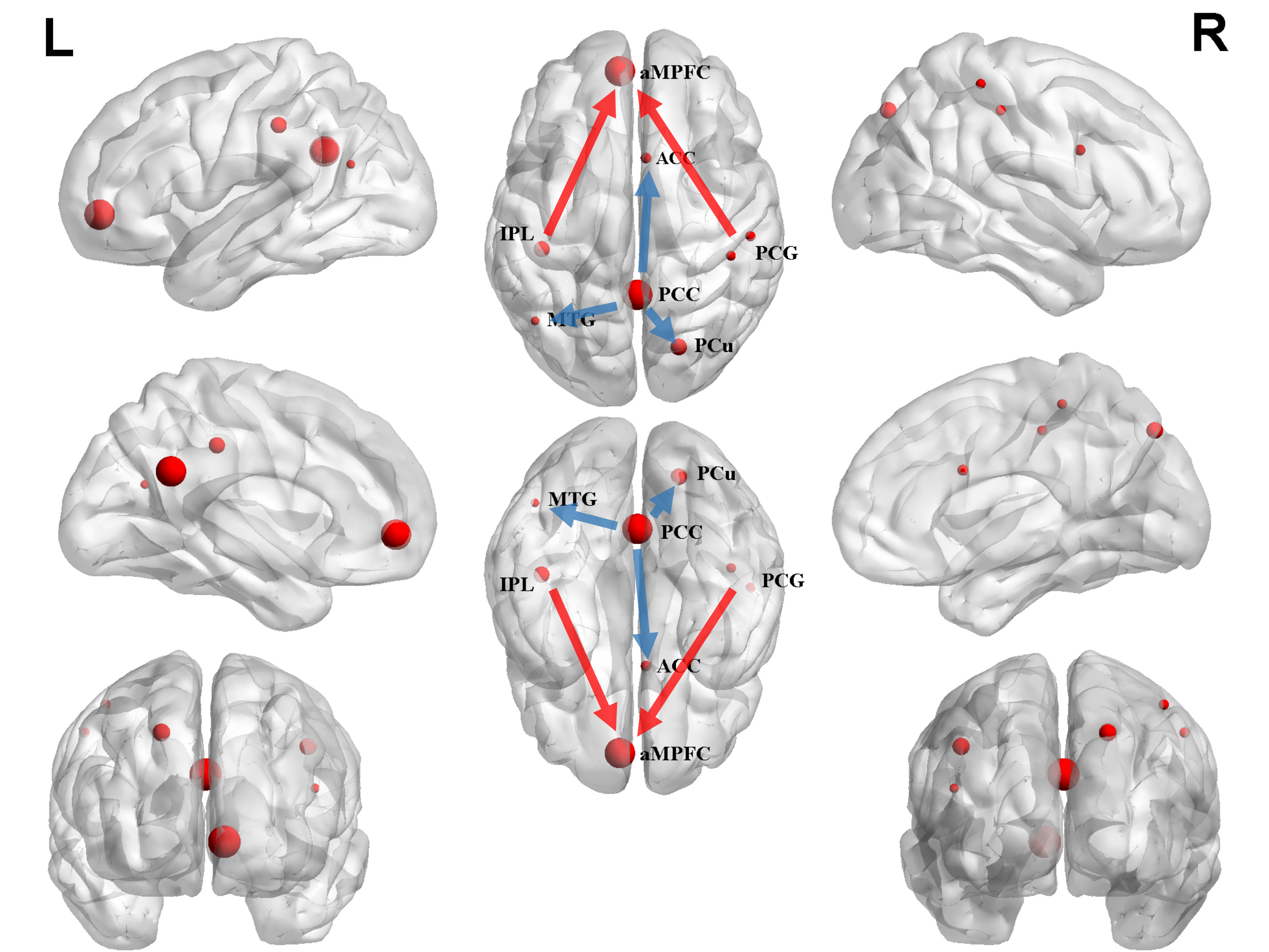
Sketch map of the between-group differences in causal connectivity: non-carriers VS. APOE ε4 carriers. The arrow of the significant causal paths represents the direction of the information flow, and the thickness and spherical radius represent the strength of the causal connectivity. Seed points in the PCC (2, -51, 27) and the aMPFC (-6, 52, -2), adapted from previously published articles (Greicius et al. 2003; Andrews-Hanna et al. 2010).
The blue arrow represents reduced effective connectivity, while the red arrow indicates increased effective connectivity.
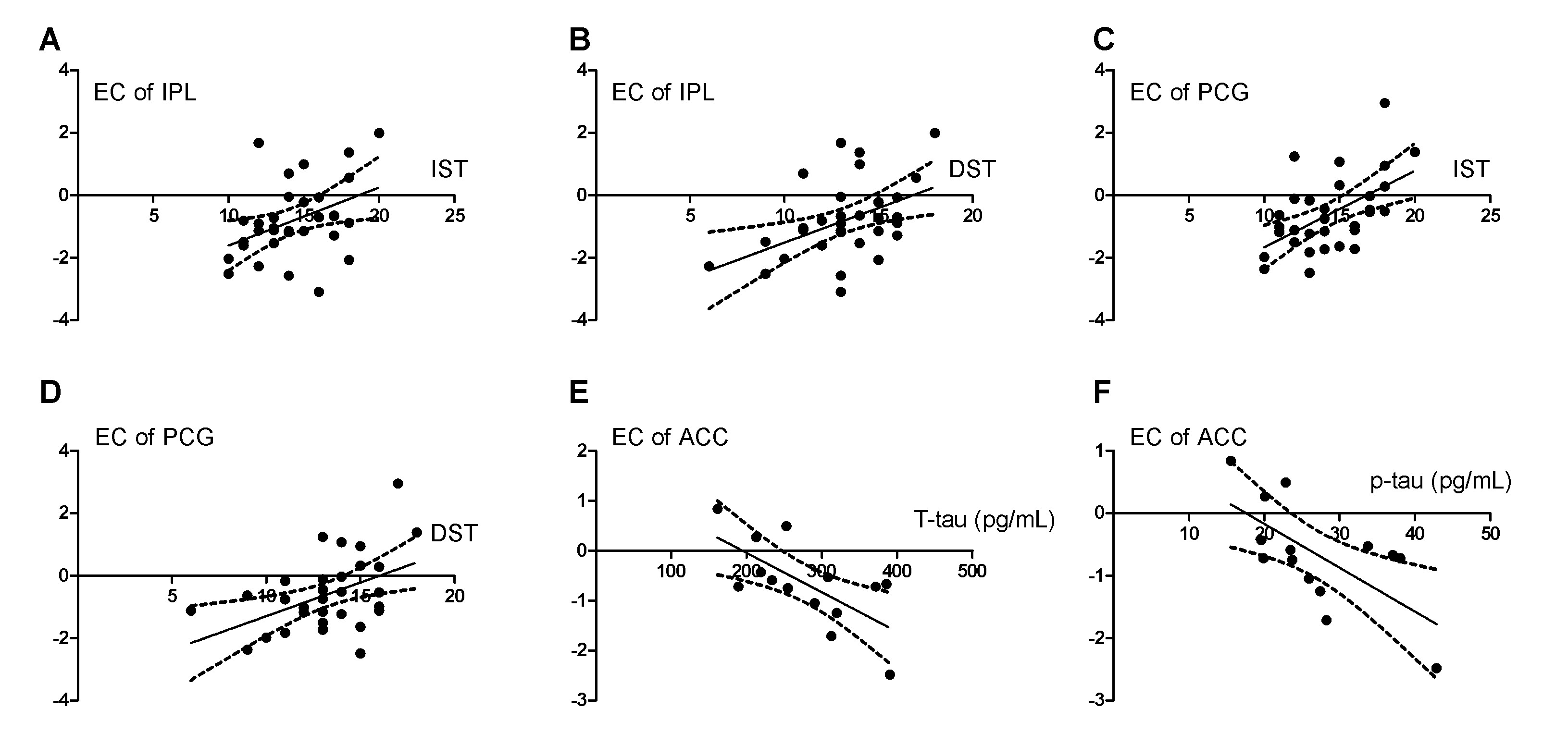
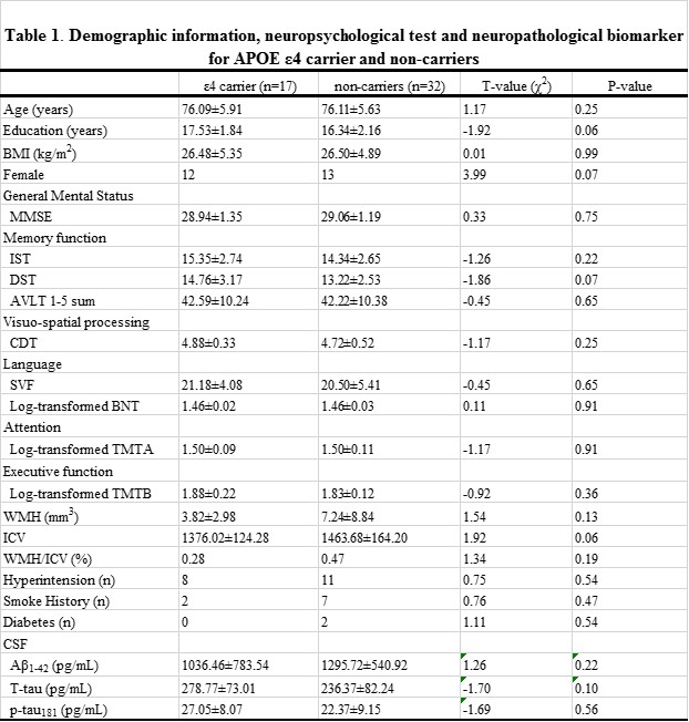
Table 1
Data is presented as means ± standard deviations.
Abbreviation: MMSE, Mini-Mental State Examination; IST, Immediate Story Recall; DST, Delayed Story Recall; AVLT, auditory verbal learning test; BNT, Boston naming test; CDT, clock drawing test; SVF, semantic verbal fluency;TMT, Trail-Making Test; ICV: Intracranial volume; WMH, white matter hyperintensities; It should be noted that mean level of Aβ1–42, t-tau and p-tau181 levels in Table only represent the subjects who had CSF sample. The final samples for CSF analyses included 28 out of 32 APOE ε4 carriers and 14 out of 17 NC