3835
Radial Simultaneous Multi Slice Imaging for Marker Tracking1Department of Radiology, Medical Physics, Medical Center ‐ University of Freiburg, Faculty of Medicine, University of Freiburg, Freiburg, Germany
Synopsis
The phase-only cross correlation (POCC) algorithm efficiently and accurately detects passive MR markers used as needle guides. In a POCC sequence the marker is automatically detected in two cross-sectional images to continuously visualize the planned needle trajectory during motion. Image acquisition of the marker, however, is very time consuming which degrades the temporal resolution. Here, it is shown that two simultaneously acquired, highly undersampled radial images - together with the consideration of the point-spread-function in the POCC algorithm - can track the marker at substantially shorter acquisition times.
Introduction
Fast and stable localization of interventional devices can be achieved with MR-safe passive markers such as needle guides for MR-guided percutaneous interventions1. The position of the marker can be reliably and effectively determined via the phase-only cross correlation (POCC) algorithm2-4 from images orthogonal to the marker’s symmetry axis. Two parallel orthogonal images are acquired to determine the direction of the marker and to align a targeting image along the marker’s symmetry axis (i.e., the theoretical needle trajectory). This way, a tracking sequence is able to provide position feedback and automatically follow the marker during a targeting procedure. As the tracking images do not contain any clinically relevant information, their acquisition times should be as short as possible to provide targeting images at a higher sampling rate5. To accelerate the tracking, we recently proposed radial acquisitions with high degree of undersampling in combination with POCC tracking that considers the projection dependent point-spread-function (PSF) of the marker6. Here, we show that this can be combined with simultaneous multi-slice imaging that does not require additional coil information.Methods
The POCC algorithm1-3 detects the position of a passive, cylindrical marker filled with a contrast agent which features a central hole for needle insertions. The marker’s position and orientation can be determined from two parallel images which are oriented perpendicular to the marker’s symmetry axis. The marker appears in a ring-like shape and can be automatically detected via a POCC algorithm. The position information allows aligning an image parallel to the marker’s symmetry axis and planning of the needle pathway. The tracking and targeting images are continuously acquired and allow following manipulations of the marker.
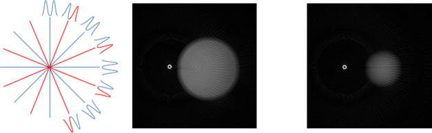
To realize faster marker tracking, we propose to use radially undersampled simultaneous multisclice (SMS) excitation7 in conjunction with a modified POCC detection algorithm for marker detection6. SMS acquisition is performed with a phase-cycled dualband pulse which leads to a constructive superposition of the first slice (in phase) and a destructive one of the second slice (out-of-phase, Fig.1, left image). To reconstruct the second slice, the phase pattern in k-space is reversed before regridding (i.e., every second radial spoke is multiplied with a phase of -1) and Fourier Transform (Fig. 1, right image).
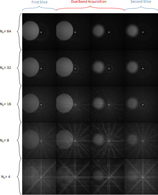
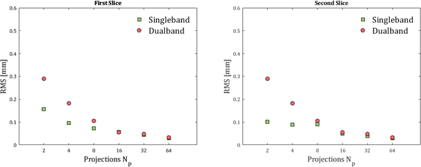
To assess the tracking accuracy of the SMS excitation, a phantom data set was acquired in a 1.5T whole body MR system (MAGNETOM Symphony, Siemens Healthcare, Erlangen, Germany) using the body array and the spine coil for reception. Images were collected perpendicular to the symmetry axis of a commercially available passive marker (Invivo GmbH, Schwerin, Germany) with different numbers of evenly spaced projections (Np = 64, 32, 16, 8, 4, 2) and for each projection number, 200 images were obtained (TR/TE = 5.64/2.20ms, α = 30°, FOV: 300×300mm², matrix: 256×256). Additionally, single band excitations were performed for comparison (Fig. 2). Reconstruction and POCC detection of the marker position was performed online with subpixel precision. The standard deviations sx and sy as well as the root-mean-square error $$$RMS = \sqrt{s_x^2+s_y^2}$$$ were calculated over each set of 200 images (Fig. 3).
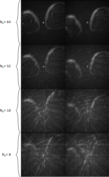
Finally, the marker was placed between the thighs of a volunteer, and a series of 200 radial SMS images of the marker cross-section was acquired. A subsequent POCC detection was performed with different numbers of projections using the same parameters as described for the phantom experiment.
Results & Discussion
Using radial SMS imaging for POCC marker detection, the marker was detected in the phantom measurements for Np ≥ 16 with comparable detection accuracy as in sequentially acquired images. Fewer Np lead to an increase of the RMS error and therefore to an unreliable marker detection accuracy which is unsuitable for clinical targeting procedures. In the in vivo experiment the marker could be detected in all images with Np≥16 projections.
Our initial study demonstrates that it is technically possible to implement simultaneously acquired radial projections for marker detection while maintaining a reasonable targeting accuracy. Compared to conventional acquisitions of the marker images, this technique allows for a 2-fold shorter acquisition time. With incorporation of this technique into the tracking sequence the marker tracking would only require very little time and might enable targeting even of moving structures.
Acknowledgements
No acknowledgement found.References
[1] Beyersdorff, D. et al. MR Imaging–guided Prostate Biopsy with a Closed MR Unit at 1.5 T: Initial Results1. Radiology 234, 576–581 (2005).
[2] de Oliveira A, et al. Automatic passive tracking of an endorectal prostate biopsy device using phase-only cross-correlation. Magn Reson Med 59:1043–1050 (2008)
[3] Krafft AJ, et al. Passive marker tracking via phase-only cross correlation (POCC) for MR-guided needle interventions: Initial in vivo experience. Physica Medica 29:607-614 (2013)
[4] Zamecnik P, et al. Automated Real-time Needle-Guide Tracking for Fast 3-T MR-guided Transrectal Prostate Biopsy: A Feasibility Study. Radiology 273:879-886 (2014)
[5] Reichert A, et al. Simultaneous slice excitation for accelerated passive marker tracking via phase-only cross correlation (POCC) in MR-guided needle interventions. MAGMA (2018)
[6] Reichert A, et al. Passive Marker Tracking with Phase-Only Cross Correlation (POCC) in Highly Undersampled Radial Images: Improvements by Point-Spread-Function Considerations. Proc. Intl. Soc. Mag. Reson. Med. 26 (2018)
[7] Yutzy SR, et al. Improvements in multislice parallel imaging using radial CAIPIRINHA. Magn. Reson. Med. 65, 1630–1637 (2011).
Figures